Copyright
©The Author(s) 2022.
World J Gastrointest Endosc. Apr 16, 2022; 14(4): 205-214
Published online Apr 16, 2022. doi: 10.4253/wjge.v14.i4.205
Published online Apr 16, 2022. doi: 10.4253/wjge.v14.i4.205
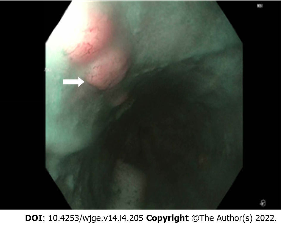
Figure 1 Endoscopy revealing skip lesions, which represent submucosal spread of the cancer in the proximal esophagus.
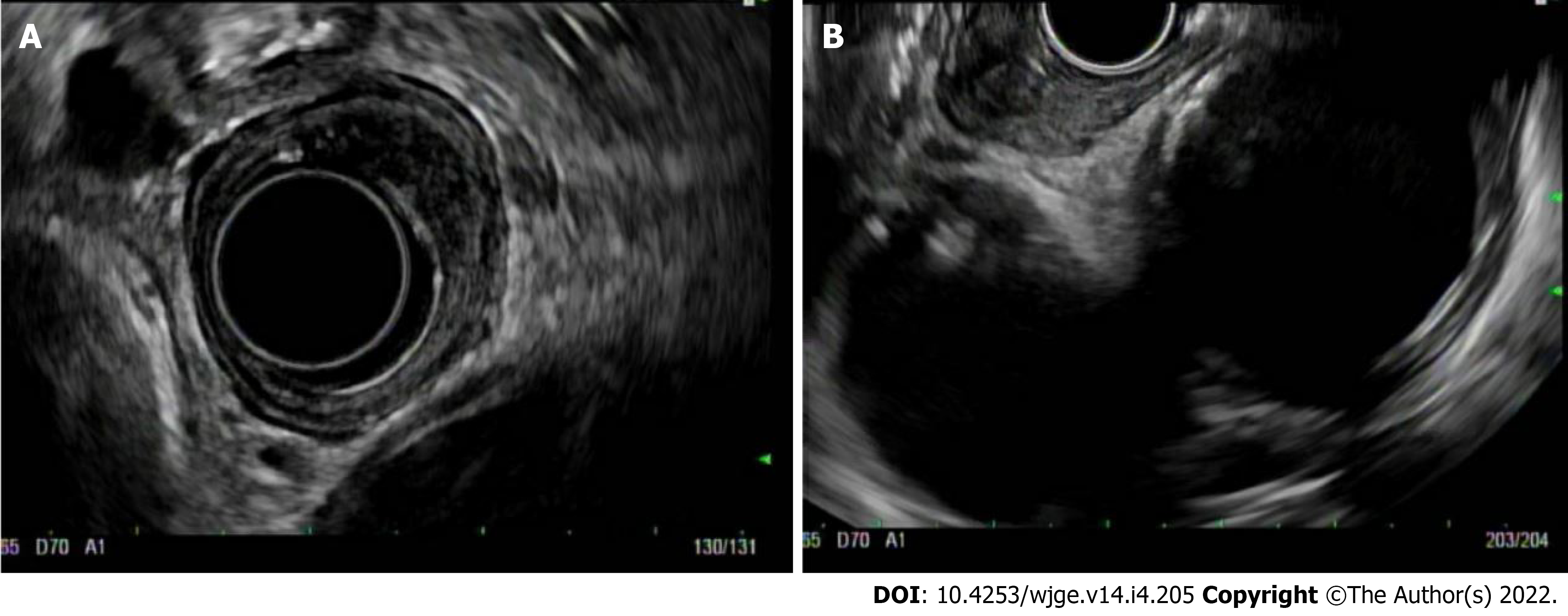
Figure 2 Radial endoscopic ultrasound view of an early esophageal cancer (A) and linear endoscopic ultrasound view of the same lesion (B).
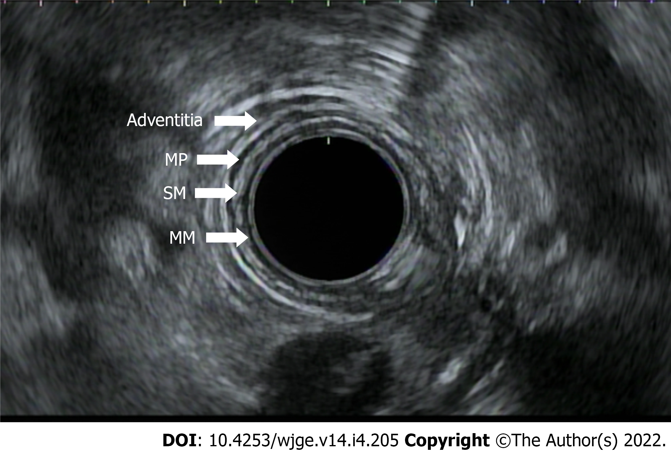
Figure 3 Endoscopic ultrasound of normal esophageal wall layers.
MM: Mucosa; SM: Submucosa; MP: Muscularis propria.
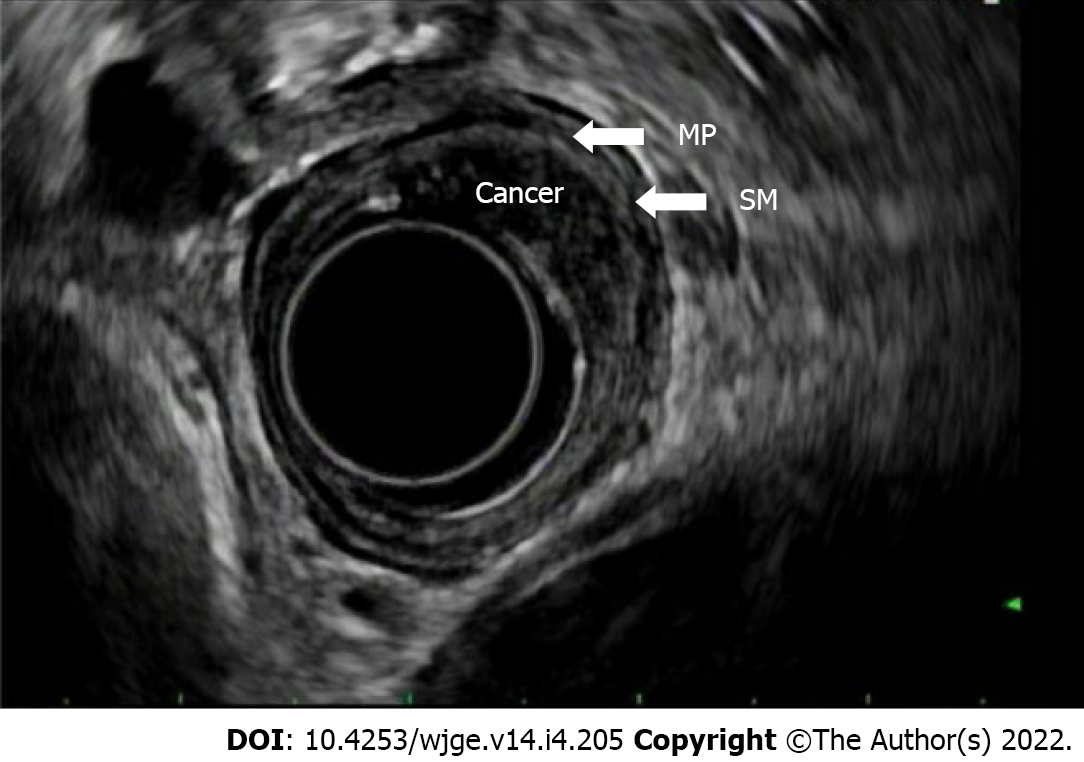
Figure 4 Endoscopic ultrasound view of a T1b esophageal cancer.
The cancer invades the submucosa but not the muscularis propria. SM: Submucosa; MP: Muscularis propria.
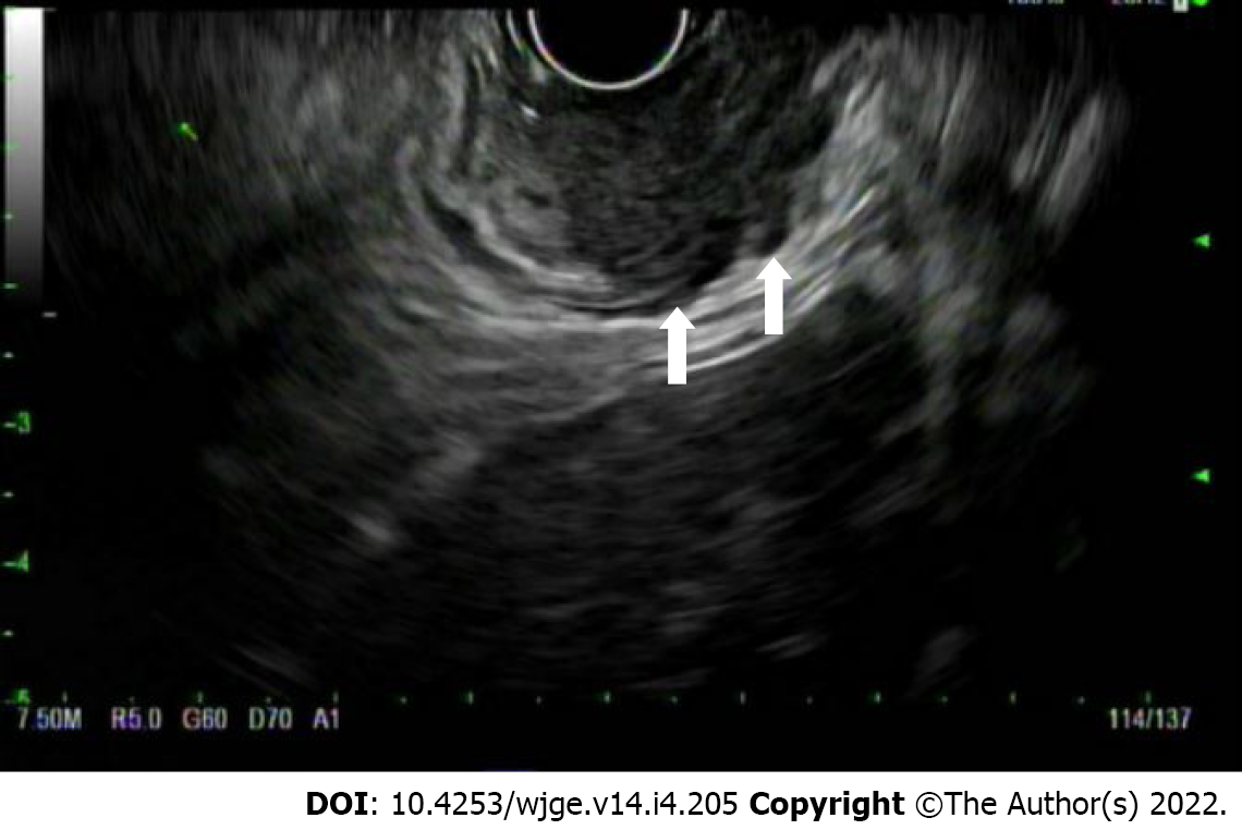
Figure 5 Endoscopic ultrasound view of a T3 esophageal cancer.
The cancer invades through the entire esophageal wall and invades the adventitia.
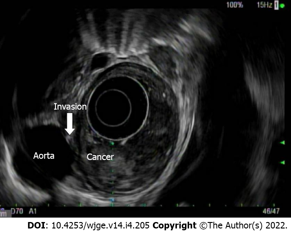
Figure 6 Endoscopic ultrasound view of a T4 esophageal cancer.
The cancer invades the aorta.
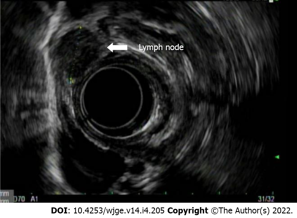
Figure 7 Endoscopic ultrasound view of a malignant peritumor lymph node.
It is hypoechoic, round, and greater than 1 cm in size and has distinct borders.
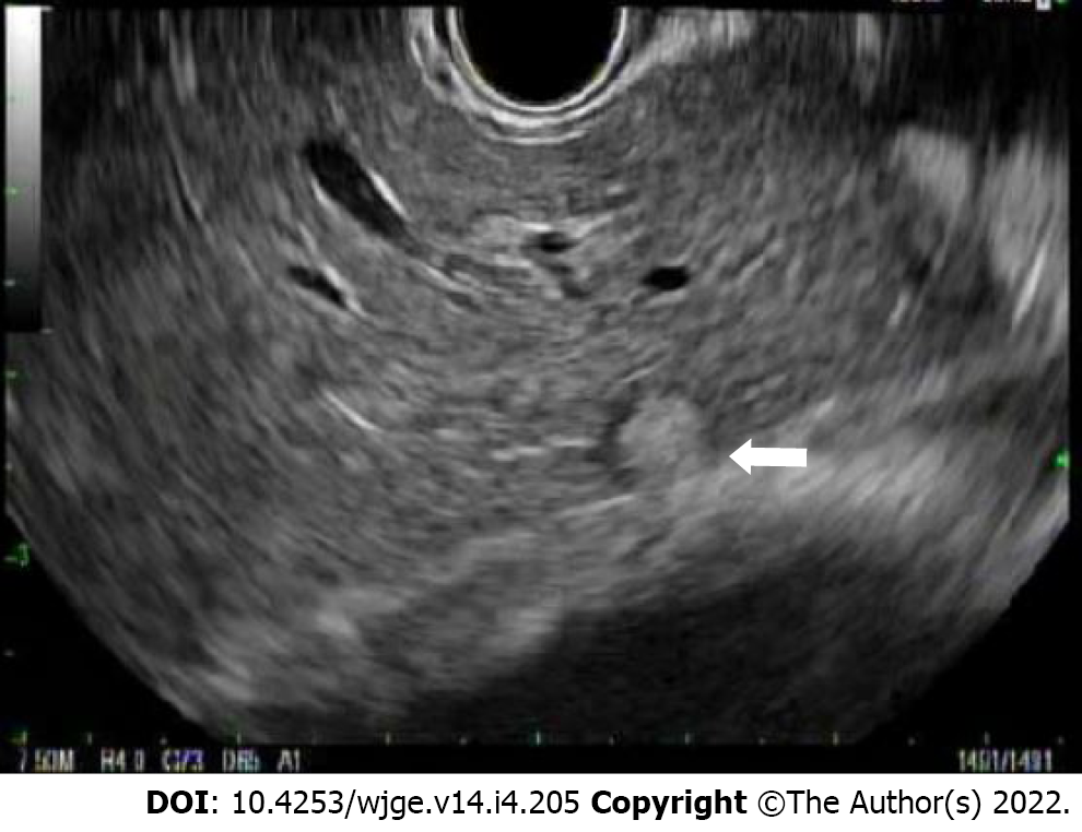
Figure 8 Endoscopic ultrasound image of a round liver metastasis.
- Citation: Radlinski M, Shami VM. Role of endoscopic ultrasound in esophageal cancer. World J Gastrointest Endosc 2022; 14(4): 205-214
- URL: https://www.wjgnet.com/1948-5190/full/v14/i4/205.htm
- DOI: https://dx.doi.org/10.4253/wjge.v14.i4.205


