Copyright
©The Author(s) 2022.
World J Gastrointest Endosc. Mar 16, 2022; 14(3): 176-182
Published online Mar 16, 2022. doi: 10.4253/wjge.v14.i3.176
Published online Mar 16, 2022. doi: 10.4253/wjge.v14.i3.176
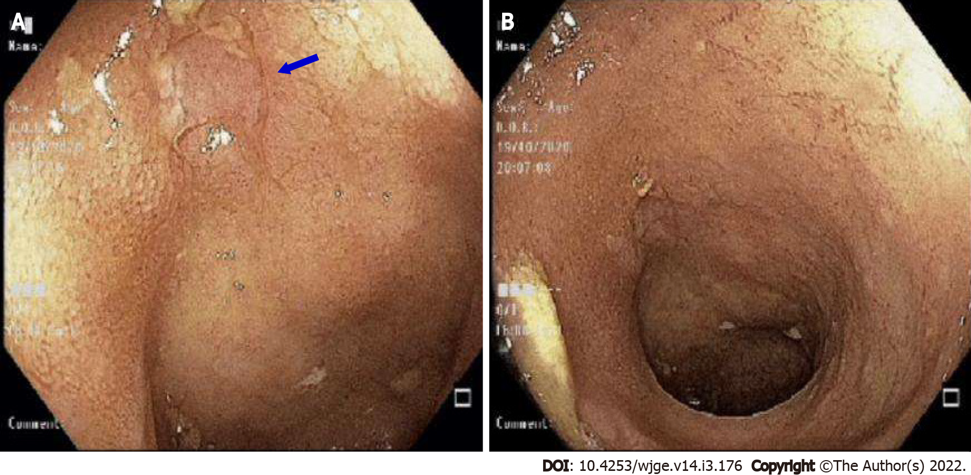
Figure 1 Colonoscopy exam.
A: Ulcer in the terminal ileum; B: Mild ulcer in terminal ileum.
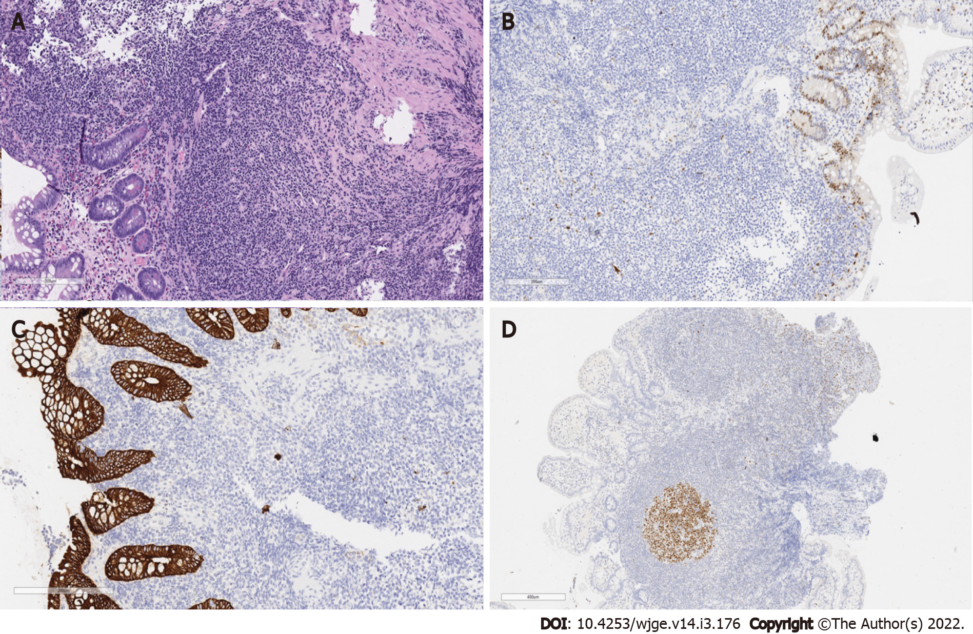
Figure 2 Biopsy of terminal ileum showing.
A: Hematoxylin & eosin-dense lymphocytic infiltrate composed of small cells, with a cytoplasmic halo, characteristic of cells in the marginal zone; B: Cyclin D1-Negative neoplastic infiltrate for protein expression of cyclin D1. Observe the positive control in the glandular epithelium; C: Cytokeratin cocktail AE1/AE3-negative neoplastic infiltrate for protein expression of cytokeratin. Observe the positive control in the glandular epithelium; D: BCL6-Protein label for BCL60. Note the negativity of the neoplastic infiltrate, and the positive internal controls in the germinal center of a reactional follicle.
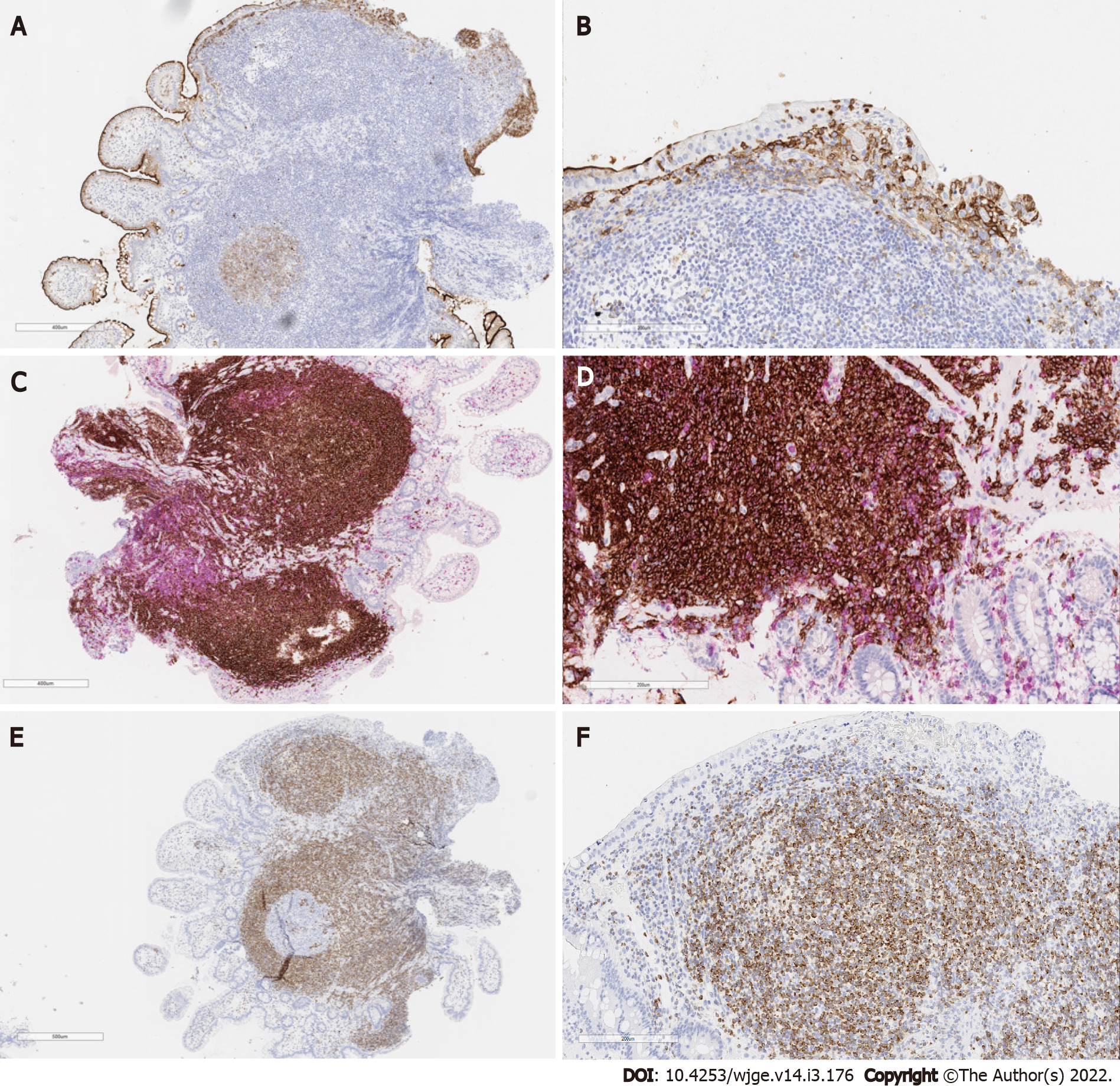
Figure 3 Terminal ileum biopsy showing.
A: Protein labeling for CD10. Note the negativity of the neoplastic infiltrate, and the positive internal controls of the glandular epithelium and germinal center of a reactional follicle; B: Protein labeling for CD10. Note the negativity of the neoplastic infiltrate and the positive internal controls of the glandular epithelium; C and D: Double labeling of CD20 (brown) and CD3 (red) demonstrating the nature of B lymphocytes of the neoplastic infiltrate. Note that T cells border the neoplastic infiltrate and preferably the epithelium, attesting to its reaction nature; E: Protein labeling for BCL2. Note that the neoplastic infiltrate expresses strongly and the internal negative control in the germinal center of a reactional follicle; F: Strong protein labeling of the neoplastic infiltrate for BCL2
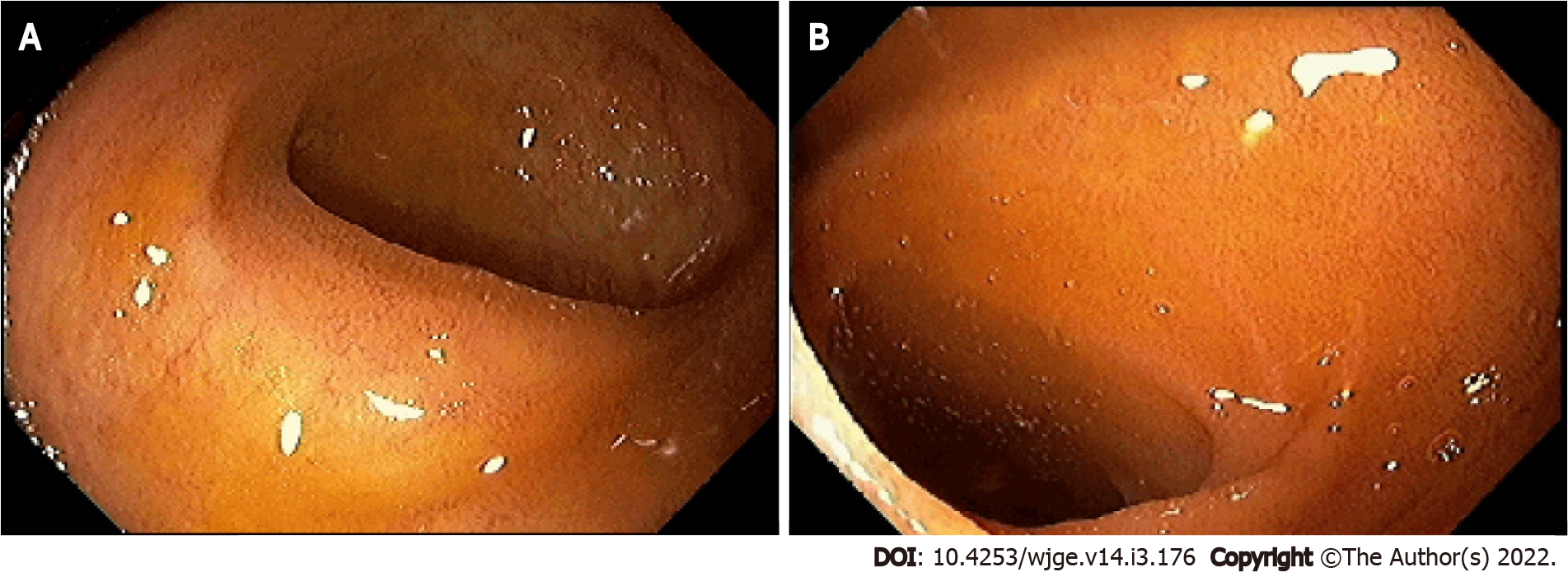
Figure 4 Colonoscopy after treatment.
A: Disease-free mucosa; B: Ulcer-free terminal ileum.
- Citation: de Figueiredo VLP, Ribeiro IB, de Moura DTH, Oliveira CC, de Moura EGH. Mucosa-associated lymphoid tissue lymphoma in the terminal ileum: A case report. World J Gastrointest Endosc 2022; 14(3): 176-182
- URL: https://www.wjgnet.com/1948-5190/full/v14/i3/176.htm
- DOI: https://dx.doi.org/10.4253/wjge.v14.i3.176


