Copyright
©The Author(s) 2022.
World J Gastrointest Endosc. Feb 16, 2022; 14(2): 96-105
Published online Feb 16, 2022. doi: 10.4253/wjge.v14.i2.96
Published online Feb 16, 2022. doi: 10.4253/wjge.v14.i2.96
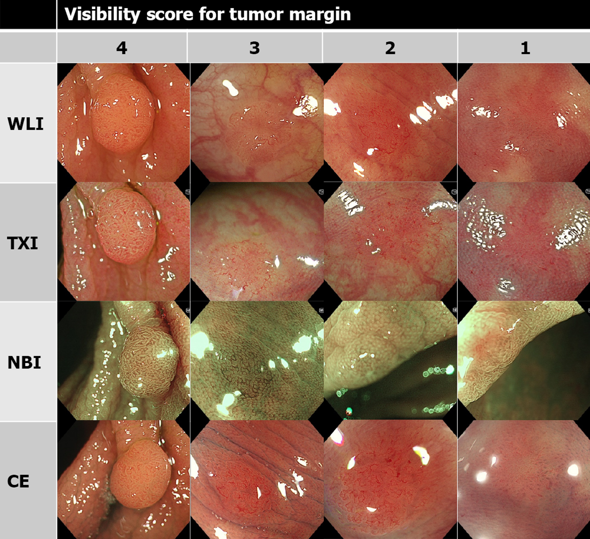
Figure 1 Representative images of visibility score for tumor margin.
Visibility score was defined as following: score 4, excellent (easily detectable); score 3, good (detectable with careful observation); score 2, fair (hardly detectable without careful examination); score 1, poor (not detectable without repeated careful examination). WLI: White light imaging; TXI: Texture and color enhancement imaging; NBI: Narrow band imaging; CE: Chromoendoscopy.
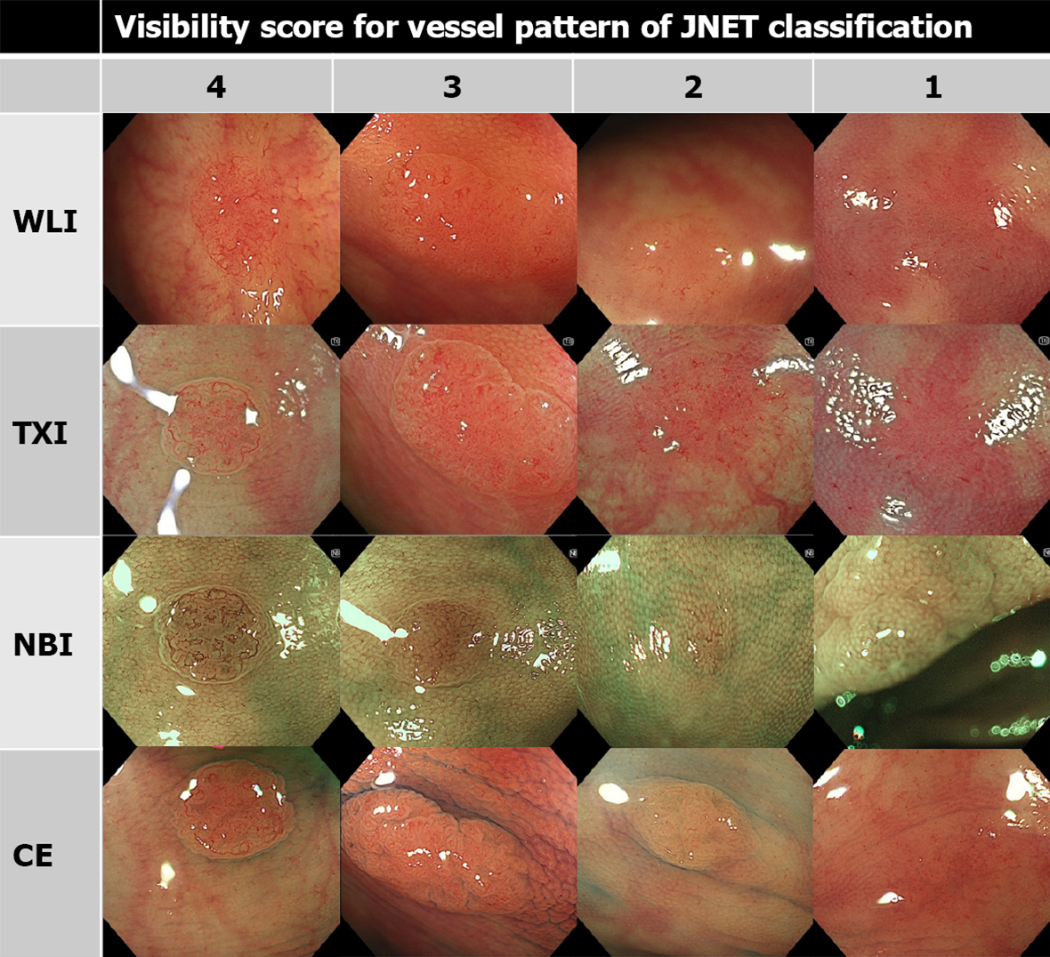
Figure 2 Representative images of visibility score for vessel pattern of Japan narrow band imaging Expert Team classification.
Visibility score was defined as following: score 4, excellent (easily detectable); score 3, good (detectable with careful observation); score 2, fair (hardly detectable without careful examination); score 1, poor (not detectable without repeated careful examination). NBI: Narrow band imaging; JNET: Japan NBI Expert Team; WLI: White light imaging; TXI: Texture and color enhancement imaging; CE: Chromoendoscopy.
Figure 3 Representative images of visibility score for surface pattern of Japan narrow band imaging Expert Team classification.
Visibility score was defined as following: score 4, excellent (easily detectable); score 3, good (detectable with careful observation); score 2, fair (hardly detectable without careful examination); score 1, poor (not detectable without repeated careful examination). NBI: Narrow band imaging; JNET: Japan NBI Expert Team; WLI: White light imaging; TXI: Texture and color enhancement imaging; CE: Chromoendoscopy.
- Citation: Toyoshima O, Nishizawa T, Yoshida S, Yamada T, Odawara N, Matsuno T, Obata M, Kurokawa K, Uekura C, Fujishiro M. Texture and color enhancement imaging in magnifying endoscopic evaluation of colorectal adenomas. World J Gastrointest Endosc 2022; 14(2): 96-105
- URL: https://www.wjgnet.com/1948-5190/full/v14/i2/96.htm
- DOI: https://dx.doi.org/10.4253/wjge.v14.i2.96


