Copyright
©The Author(s) 2022.
World J Gastrointest Endosc. Dec 16, 2022; 14(12): 739-747
Published online Dec 16, 2022. doi: 10.4253/wjge.v14.i12.739
Published online Dec 16, 2022. doi: 10.4253/wjge.v14.i12.739
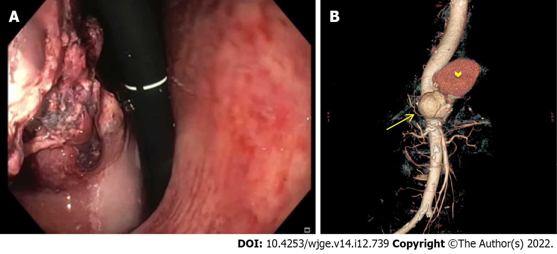
Figure 1 Severe non-variceal upper gastrointestinal bleeding due to primary aorto-gastric fistula.
A: Retroflexed endoscopic view showing gastric bulging mass partially covered by blood clots, originating from the fundus and extending to the posterior wall of the proximal body; B: Three-dimensional computed tomography angiography showing ruptured thoracoabdominal aortic aneurysm (arrow), retained by a periaortic hematoma (arrowhead).
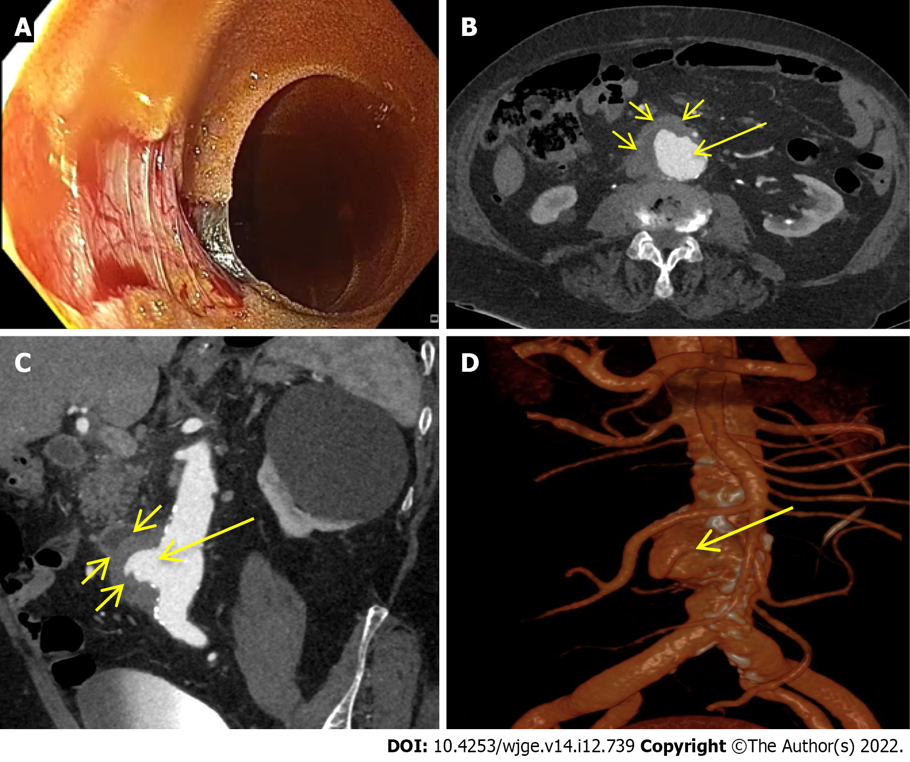
Figure 2 Severe non-variceal upper gastrointestinal bleeding due to primary aorto-duodenal fistula.
A: Esophagogastroduodenoscopy showing a large pulsating wall defect of the third duodenal portion; B-D: Axial computed tomography artery phase (B), coronal-oblique maximum intensity projection artery phase (C) and three-dimensional volume rendering reconstruction (D) showing a large outpouching from the right anterolateral wall of the abdominal aorta (B-D; long arrow) at the level of the third duodenal portion with loss of interface fat plane (B and C; short arrows), in the absence of neither air bubble within the aortic lumen and wall nor contrast medium extravasation into the duodenal lumen.
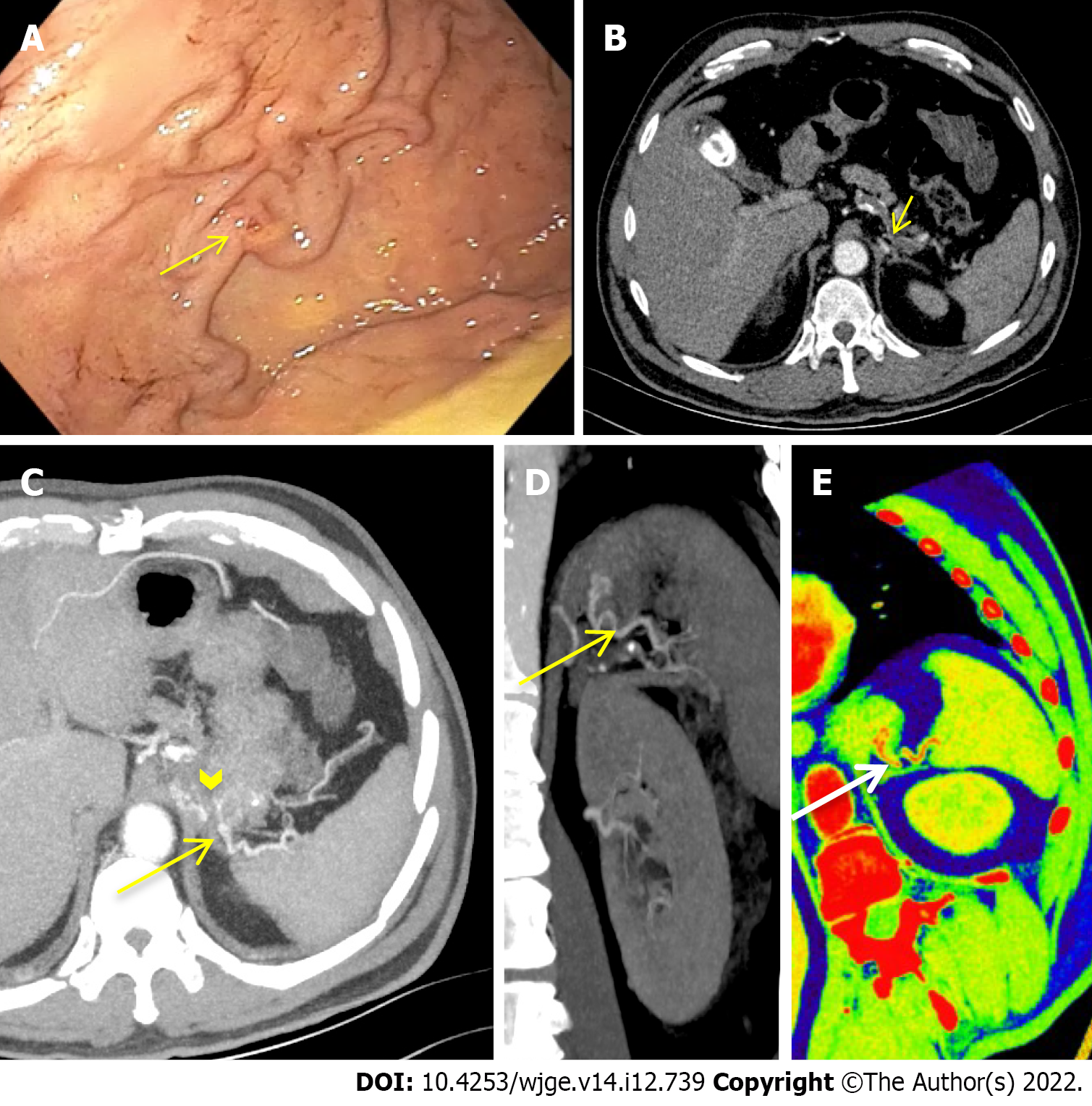
Figure 3 Severe non variceal upper gastrointestinal bleeding due to gastric submucosal arterial collaterals secondary to splenic artery thrombosis.
A: Retroflexed endoscopic view of the gastric fundus showing varicose-shaped submucosal vessels with a small erosion (arrow); B-E: Axial computed tomography dual-energy arterial phase (B) with maximum intensity projection artery phase reconstruction on axial (C) and coronal (D) multiplanar view and oblique-coronal colorimetric low keV (E) showing splenic artery thrombosis (B: short arrow) with an arterial cluster at the gastric fundus (C: arrowhead) arising from splenic artery collateral vessels (C-E: long arrow).
- Citation: Martino A, Di Serafino M, Amitrano L, Orsini L, Pietrini L, Martino R, Menchise A, Pignata L, Romano L, Lombardi G. Role of multidetector computed tomography angiography in non-variceal upper gastrointestinal bleeding: A comprehensive review. World J Gastrointest Endosc 2022; 14(12): 739-747
- URL: https://www.wjgnet.com/1948-5190/full/v14/i12/739.htm
- DOI: https://dx.doi.org/10.4253/wjge.v14.i12.739


