Copyright
©The Author(s) 2022.
World J Gastrointest Endosc. Oct 16, 2022; 14(10): 648-656
Published online Oct 16, 2022. doi: 10.4253/wjge.v14.i10.648
Published online Oct 16, 2022. doi: 10.4253/wjge.v14.i10.648
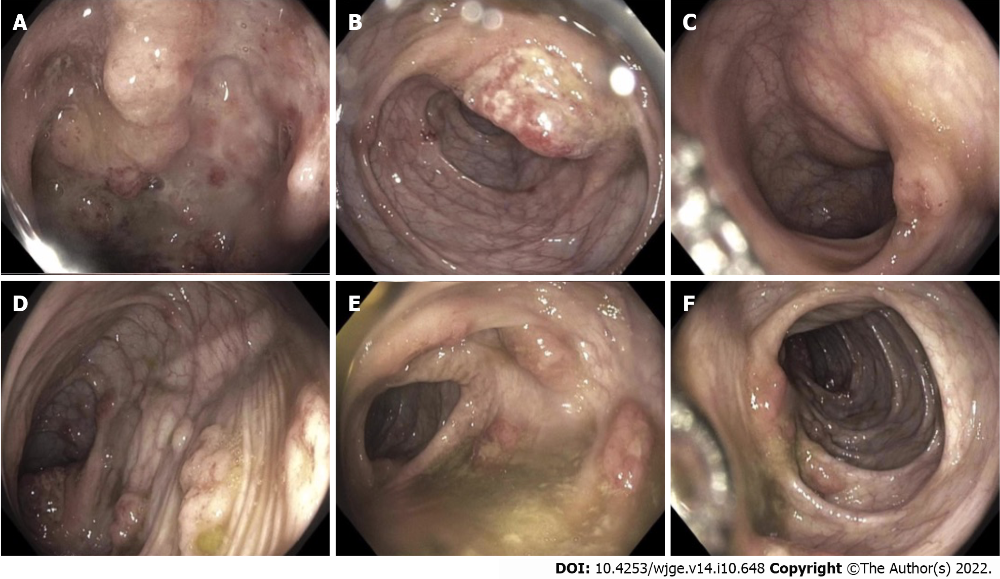
Figure 1 Colonoscopy findings.
Diffuse and severe inflammation characterized by mucosal edema, erythema, friability, pseudopolyps, and serpentine ulcerations. A: Terminal ileum; B: Ileocecal valve; C: Transverse colon; D and E: Descending colon; F: Ascending colon.
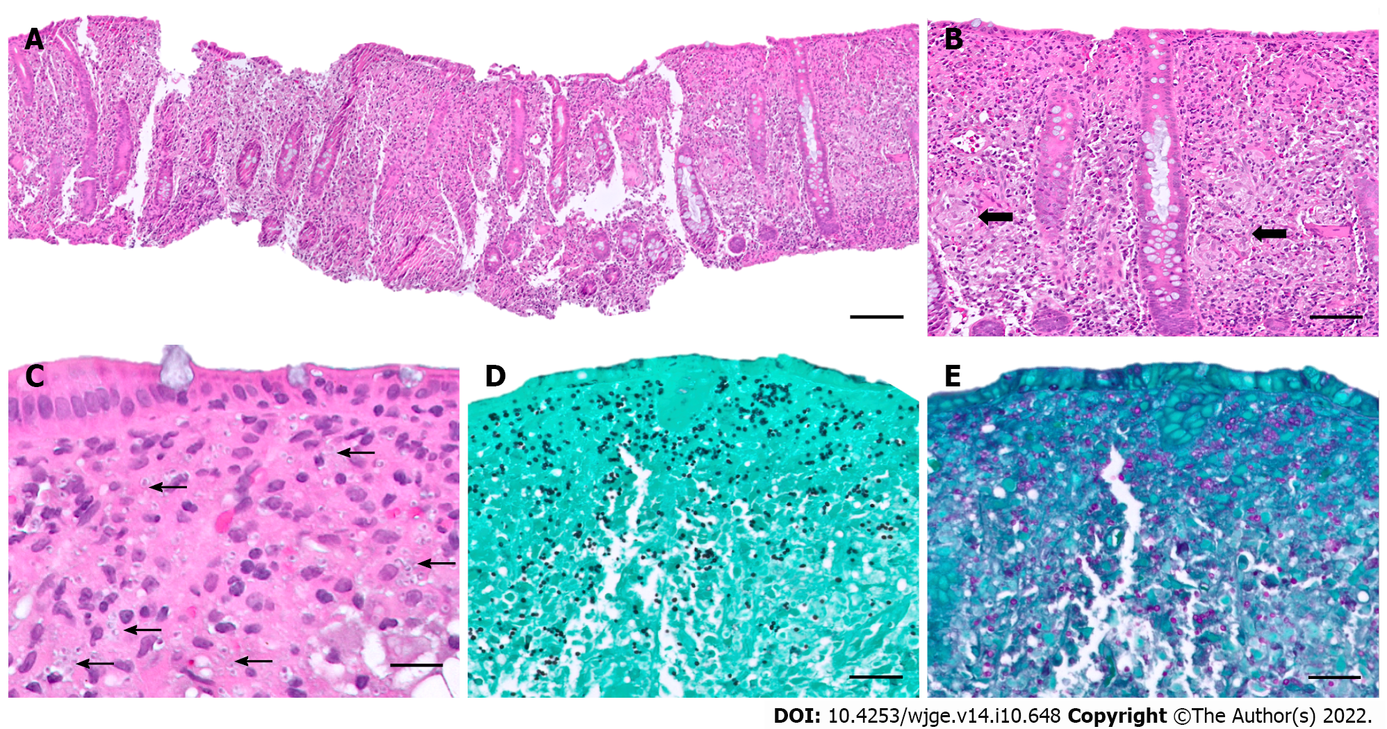
Figure 2 Histologic findings.
A: Colon biopsy revealed diffuse cellular infiltrate within the lamina propria (hematoxlyin and eosin, × 2, scale bar 1 mm); B: Scattered poorly formed granulomas (arrows) (hematoxlyin and eosin, × 20, scale bar 100 μm); C: Intracellular microorganisms (arrows) (hematoxlyin and eosin, × 40, scale bar 50 μm); Numerous yeast forms suggestive of Histoplasma spp. confirmed by special stains; D: Grocott-Gomori’s Methenamine Silver stain (× 20, scale bar 100 μm); E: Periodic acid Schiff stain (× 20, scale bar 100 μm).
- Citation: Miller CQ, Saeed OAM, Collins K. Gastrointestinal histoplasmosis complicating pediatric Crohn disease: A case report and review of literature. World J Gastrointest Endosc 2022; 14(10): 648-656
- URL: https://www.wjgnet.com/1948-5190/full/v14/i10/648.htm
- DOI: https://dx.doi.org/10.4253/wjge.v14.i10.648


