Copyright
©The Author(s) 2021.
World J Gastrointest Endosc. Sep 16, 2021; 13(9): 426-436
Published online Sep 16, 2021. doi: 10.4253/wjge.v13.i9.426
Published online Sep 16, 2021. doi: 10.4253/wjge.v13.i9.426
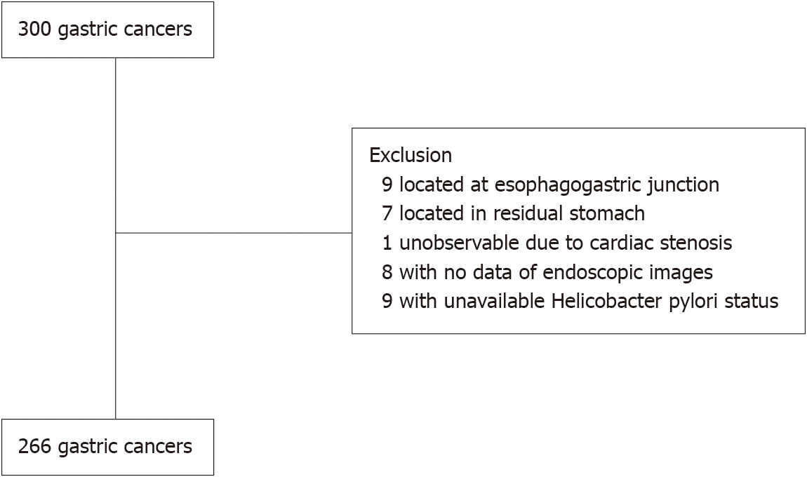
Figure 1
Patient flowchart.
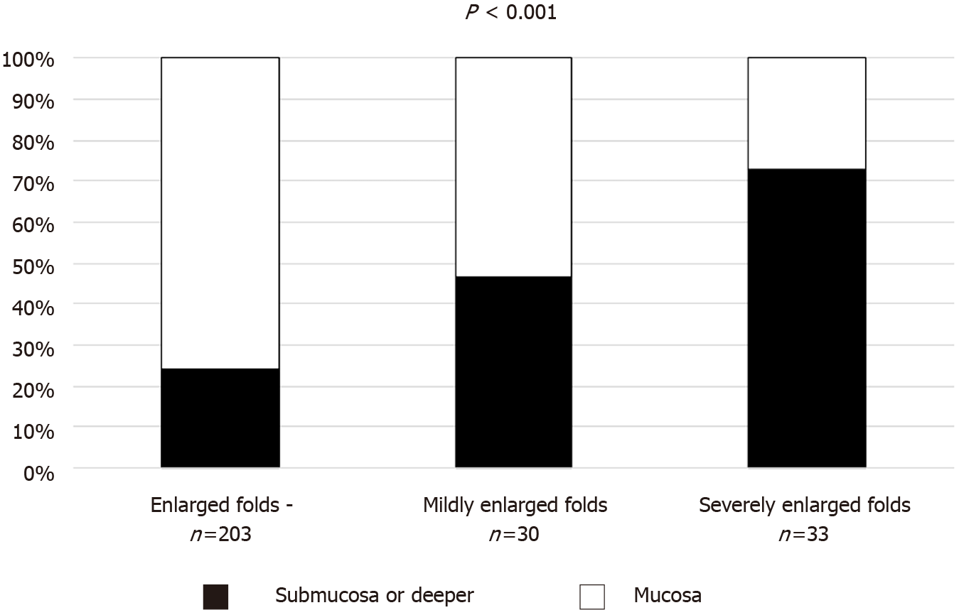
Figure 2 Proportion of submucosal invasion based on severity of enlarged folds.
The P value was calculated using the Cochran-Armitage trend test.
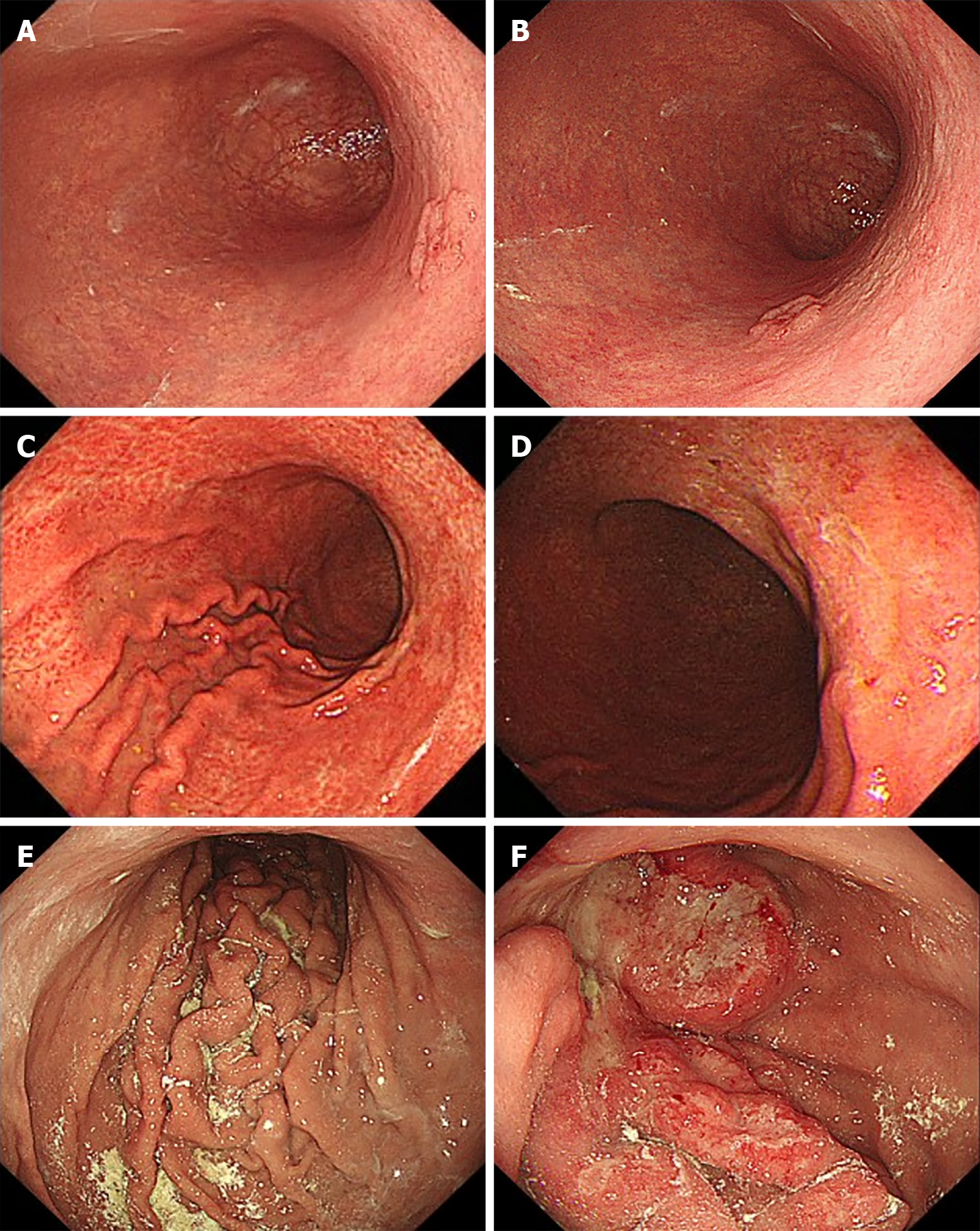
Figure 3 Representative images of enlarged folds and coexisting gastric cancer.
A and B: Enlarged fold-negative; 74-year-old man with current Helicobacter pylori (H. pylori) infection. The cancer was limited to the mucosa and was intestinal-type; C and D: Mildly enlarged folds; 40-year-old woman with current H. pylori infection. The cancer invaded the submucosa and was diffuse-type; E and F: Severely enlarged folds; 60-year-old man with current H. pylori infection. The cancer invaded the serosa and was diffuse-type. A, C and E: Greater curvature of the body; B, D and F: Gastric cancer.
- Citation: Toyoshima O, Yoshida S, Nishizawa T, Toyoshima A, Sakitani K, Matsuno T, Yamada T, Matsuo T, Nakagawa H, Koike K. Enlarged folds on endoscopic gastritis as a predictor for submucosal invasion of gastric cancers. World J Gastrointest Endosc 2021; 13(9): 426-436
- URL: https://www.wjgnet.com/1948-5190/full/v13/i9/426.htm
- DOI: https://dx.doi.org/10.4253/wjge.v13.i9.426


