Copyright
©The Author(s) 2021.
World J Gastrointest Endosc. Sep 16, 2021; 13(9): 382-390
Published online Sep 16, 2021. doi: 10.4253/wjge.v13.i9.382
Published online Sep 16, 2021. doi: 10.4253/wjge.v13.i9.382
Figure 1 Endoscopic appearance.
A: Endoscopic appearance of a Crohn’s disease fibrostenotic lesion in the ileocecal valve; B: Wire-guided 18 mm balloon dilation catheter (CRE PRO, Boston Scientific, Marlborough, MA, United States); C: Appearance after dilation.
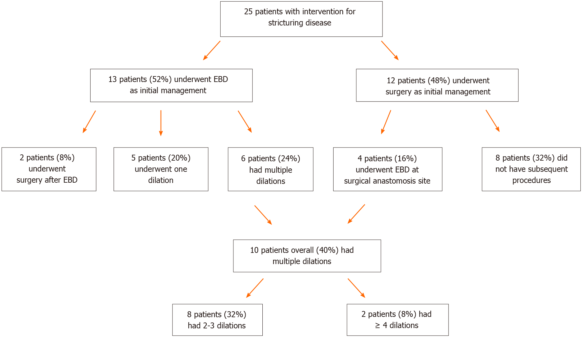
Figure 2 Management of patients with stricturing Crohn’s disease via surgery or endoscopic balloon dilation.
EBD: Endoscopic balloon dilation.
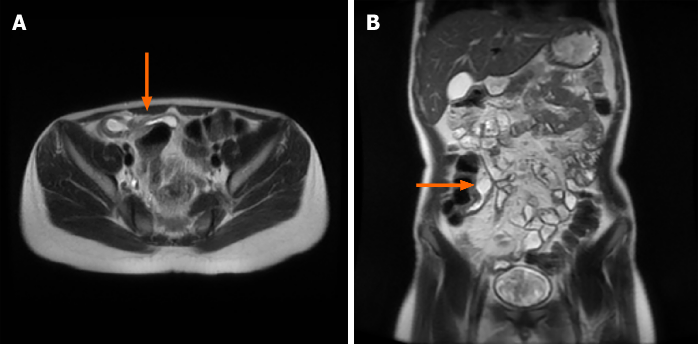
Figure 3 Magnetic resonance imaging of fibrostenosing Crohn’s disease.
A: Cross sectional magnetic resonance imaging showing the lesion in the distal ileum; B: Coronal cut on magnetic resonance imaging of fibrostenosing Crohn’s disease with proximal dilation.
- Citation: McSorley B, Cina RA, Jump C, Palmadottir J, Quiros JA. Endoscopic balloon dilation for management of stricturing Crohn’s disease in children. World J Gastrointest Endosc 2021; 13(9): 382-390
- URL: https://www.wjgnet.com/1948-5190/full/v13/i9/382.htm
- DOI: https://dx.doi.org/10.4253/wjge.v13.i9.382


