Copyright
©The Author(s) 2021.
World J Gastrointest Endosc. Dec 16, 2021; 13(12): 607-618
Published online Dec 16, 2021. doi: 10.4253/wjge.v13.i12.607
Published online Dec 16, 2021. doi: 10.4253/wjge.v13.i12.607
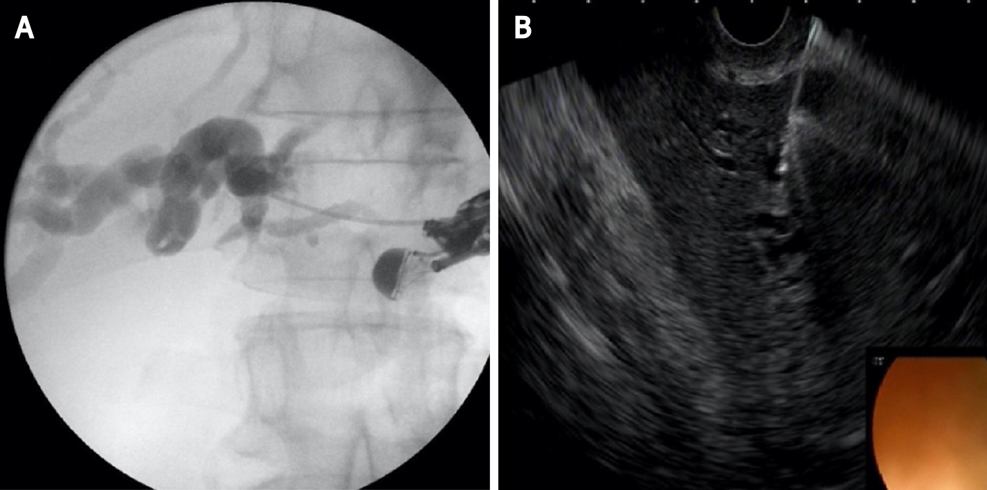
Figure 1 Left hepatic duct puncture and contrast injection.
A: Cholangiogram; B: endoscopic ultrasound image.
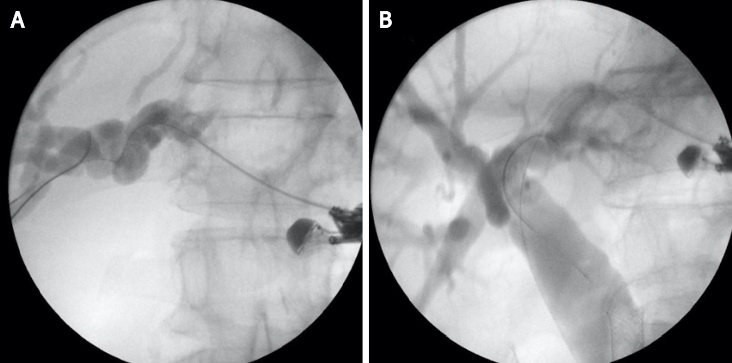
Figure 2 Hydrophilic guidewire insertion.
A: In left hepatic duct; B: To the distal common bile duct.
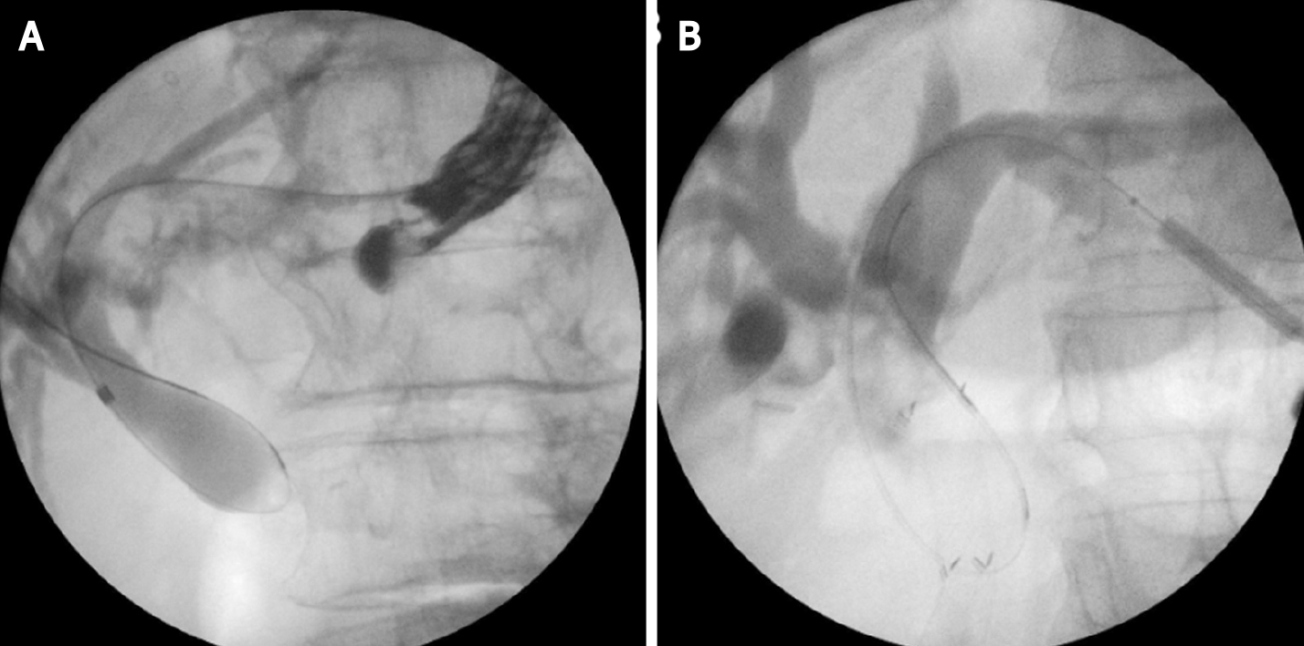
Figure 3 Tract dilatation.
A: Biliary dilation catheter; B: 4 mm balloon dilatator.
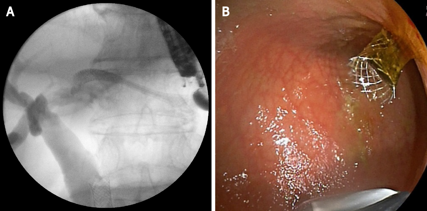
Figure 4 Stent placement-self-expandable metal stent.
A: Cholangiogram; B: Endoscopic image.
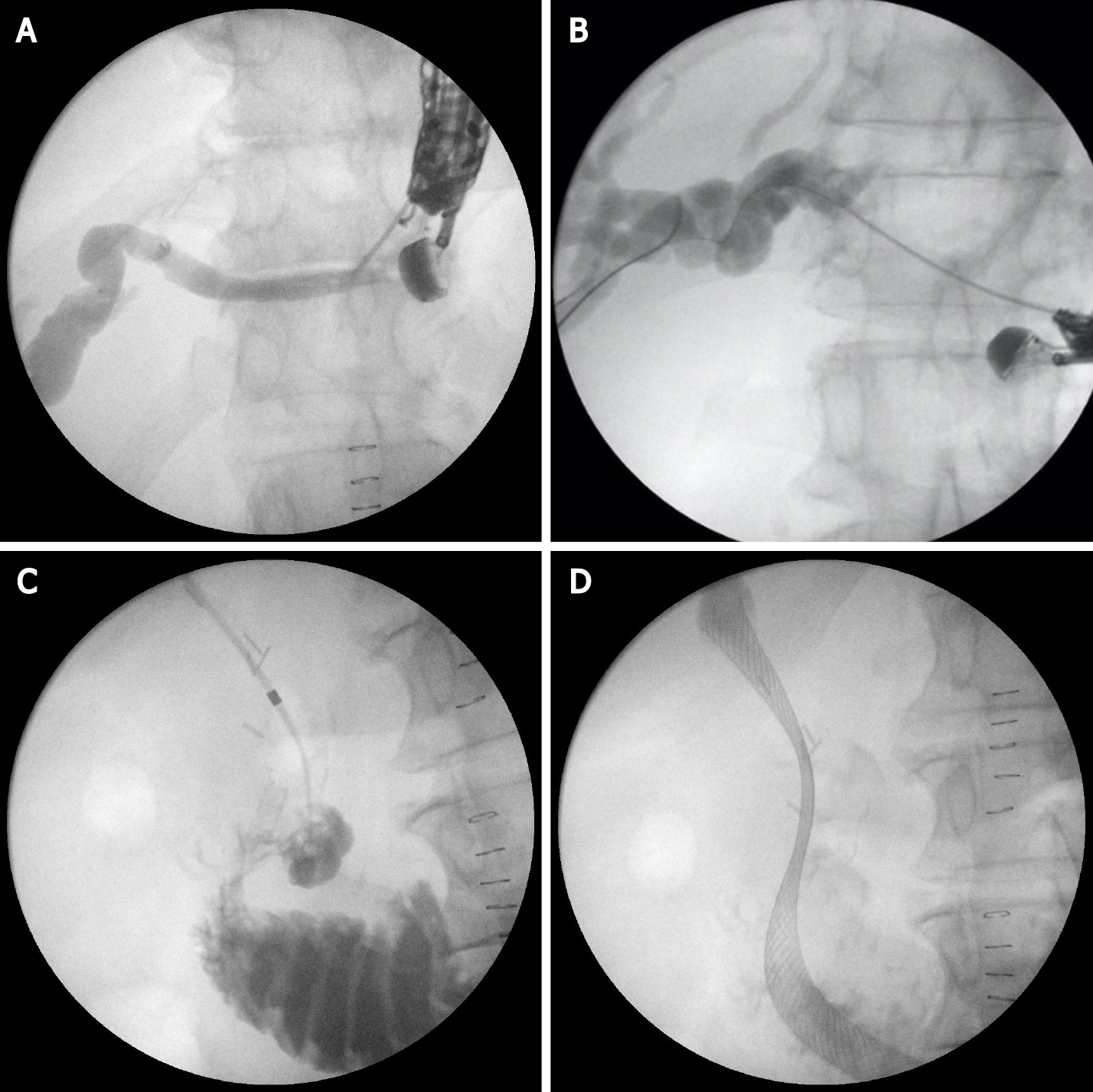
Figure 5 Endoscopic ultrasound-guided antegrade stenting.
A: Left hepatic duct puncture with 19G needle; B: Guide-wire insertion; C: Tract dilatation and advancing the biliary catheter tip transpapillary in the duodenum; D: Self-expandable metal stent placement.
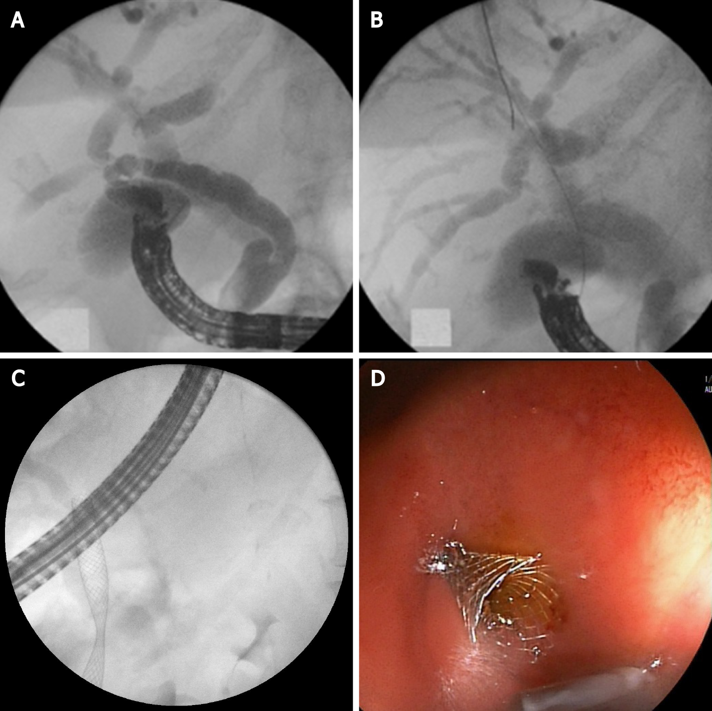
Figure 6 Endoscopic ultrasound-guided choledochoduodenostomy.
A: Puncture of the common bile duct with 19G needle and contrast injection; B: Hydrophilic guidewire inserted through the needle into bile ducts; C: Fluoroscopic image of self-expandable metal stent (SEMS); D: Endoscopic image of SEMS.
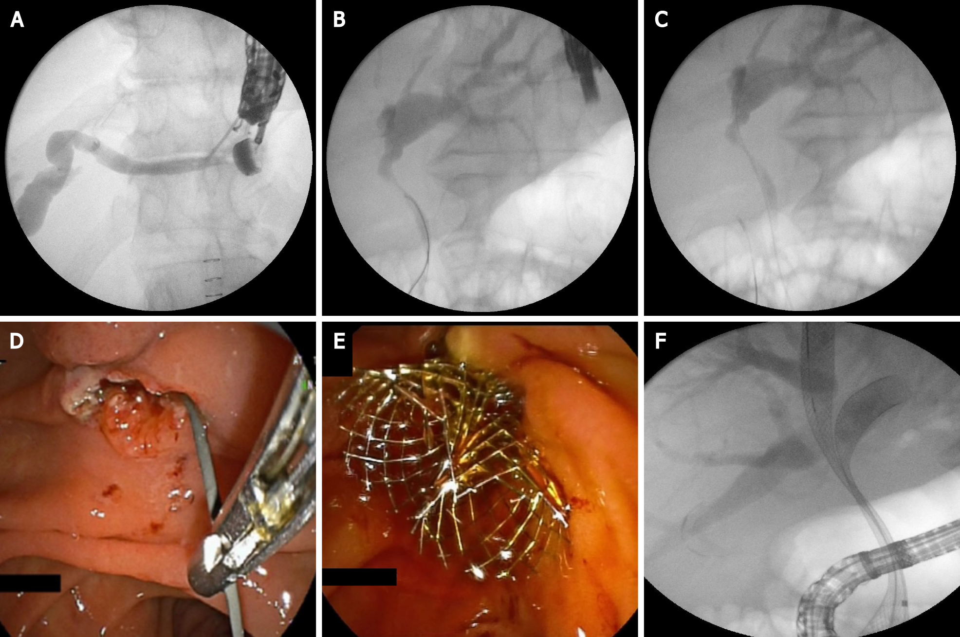
Figure 7 Endoscopic ultrasound-guided rendezvous technique.
A: Puncture the left hepatic duct with 19G needle; B: Guide-wire insertion in bile ducts; C: Guide-wire insertion transpapillary in the duodenum; D: Grasping the guide-wire with a rath tooth forceps; E: Endoscopic image of two self-expandable metal stent (SEMS); F: Fluoroscopic image of two SEMS.
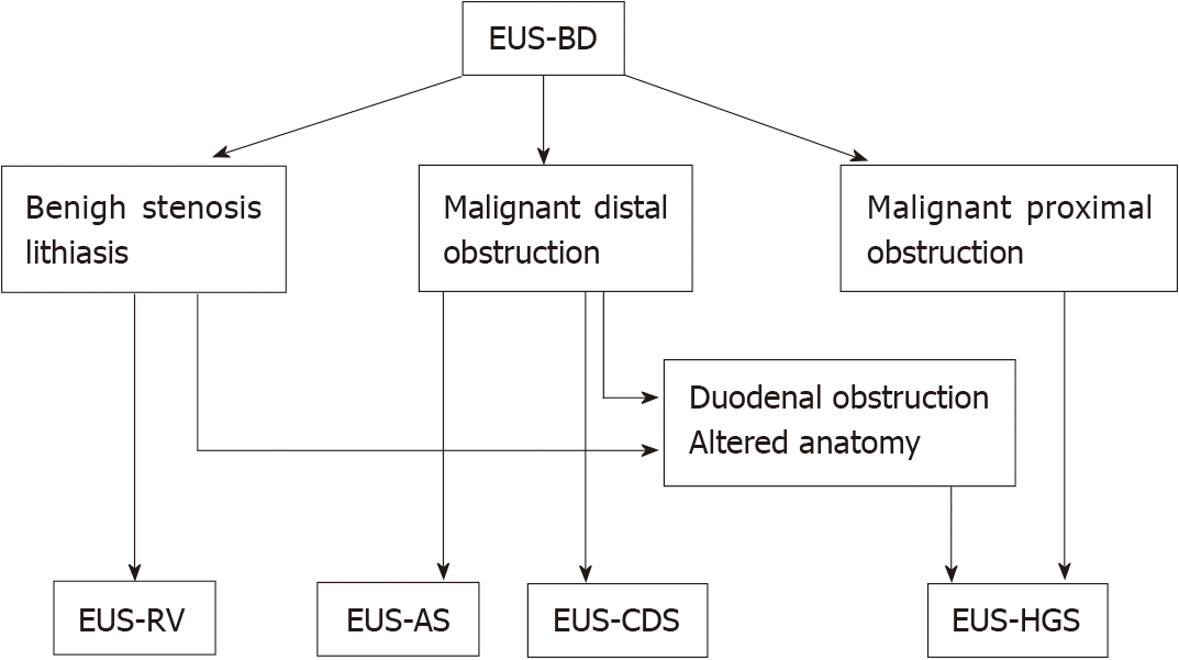
Figure 8 Current place of endoscopic ultrasound-guided biliary drainage in endoscopic biliary drainage therapy.
EUS-BD: Endoscopic ultrasound-guided biliary drainage; EUS-RV: EUS-guided rendezvous technique; EUS-AS: EUS-guided antegrade stenting; EUS-CDS: EUS-guided choledochoduodenostomy/choledochoantrostomy; EUS-HGS: EUS-guided hepaticogastrostomy.
- Citation: Karagyozov PI, Tishkov I, Boeva I, Draganov K. Endoscopic ultrasound-guided biliary drainage-current status and future perspectives. World J Gastrointest Endosc 2021; 13(12): 607-618
- URL: https://www.wjgnet.com/1948-5190/full/v13/i12/607.htm
- DOI: https://dx.doi.org/10.4253/wjge.v13.i12.607


