Copyright
©The Author(s) 2021.
World J Gastrointest Endosc. Dec 16, 2021; 13(12): 593-606
Published online Dec 16, 2021. doi: 10.4253/wjge.v13.i12.593
Published online Dec 16, 2021. doi: 10.4253/wjge.v13.i12.593
Figure 1 Composite intestinal adenoma-microcarcinoid consisting of tubulovillous adenoma with high grade dysplasia and microcarcinoid components (arrowheads) at its base.
A: The overall polyp architecture is preserved (Hematoxylin and eosin, 50 ×); B: Microcarcinoid component shows bland cytology, within edematous stroma with conspicuous eosinophils, resembling desmoplasia (Hematoxylin and eosin, 200 ×).
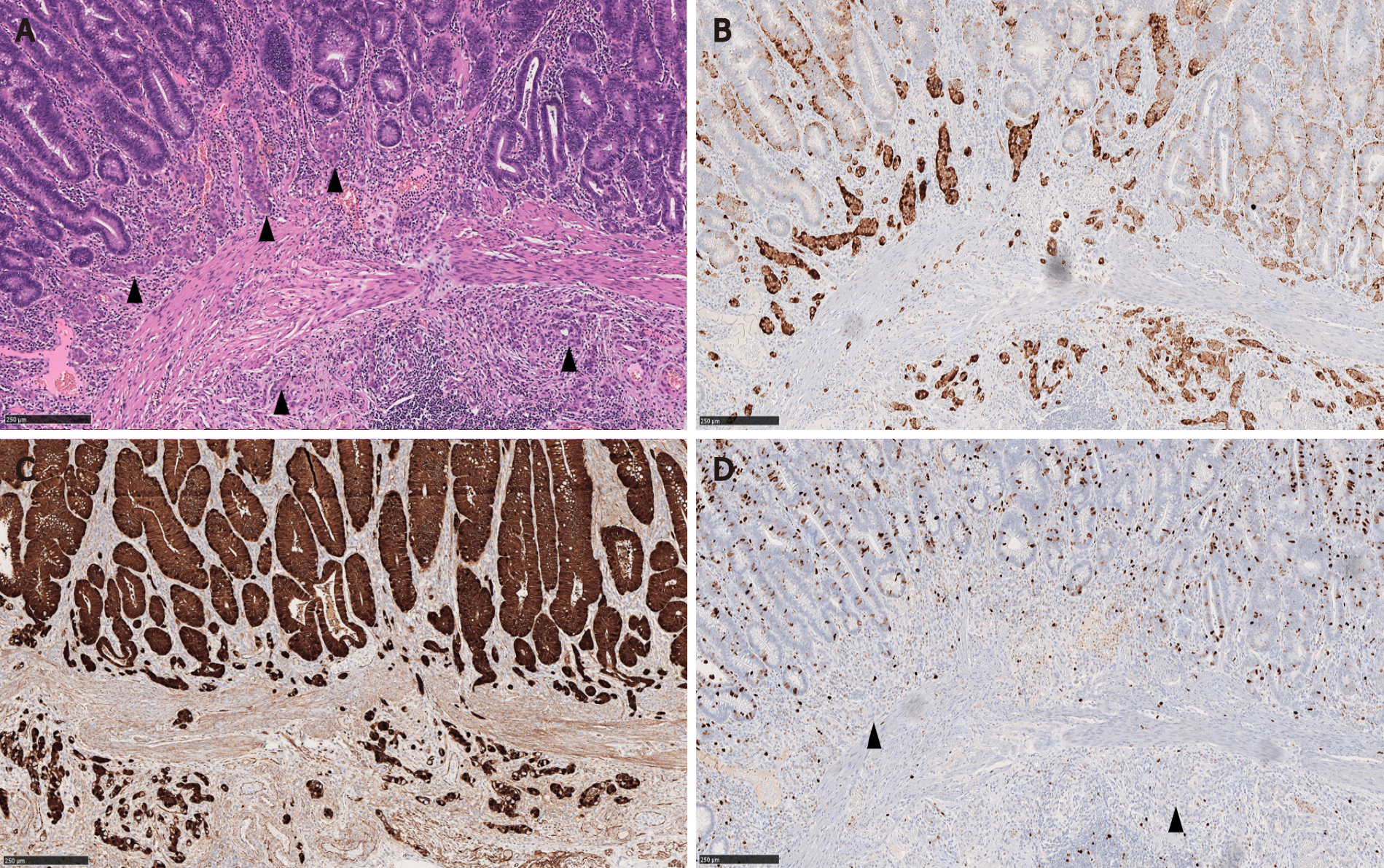
Figure 2 Composite intestinal adenoma-microcarcinoid with submucosal invasion of the microcarcinoid component.
A: The microcarcinoid (MC) components (arrowheads) form small nests that are sparsely distributed at the polyp base. No nodules or masses are grossly evident (Hematoxylin and eosin, 100 ×); B-D: The MC components are positive for synaptophysin and beta-catenin (nuclear stain) with low proliferative rate (arrowheads) (B: Synaptophysin immunostain, 100 ×; C: Beta-catenin immunostain, 100 ×; D: Ki 67 immunostain, 100 ×).
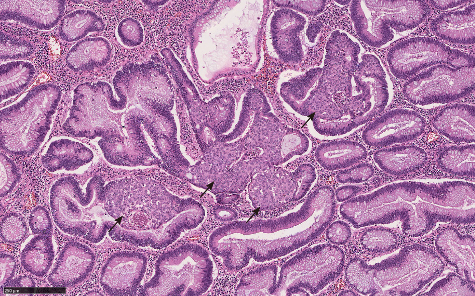
Figure 3 Squamous morules (arrows) with associated tubulovillous adenoma (Hematoxylin and eosin, 100 ×).
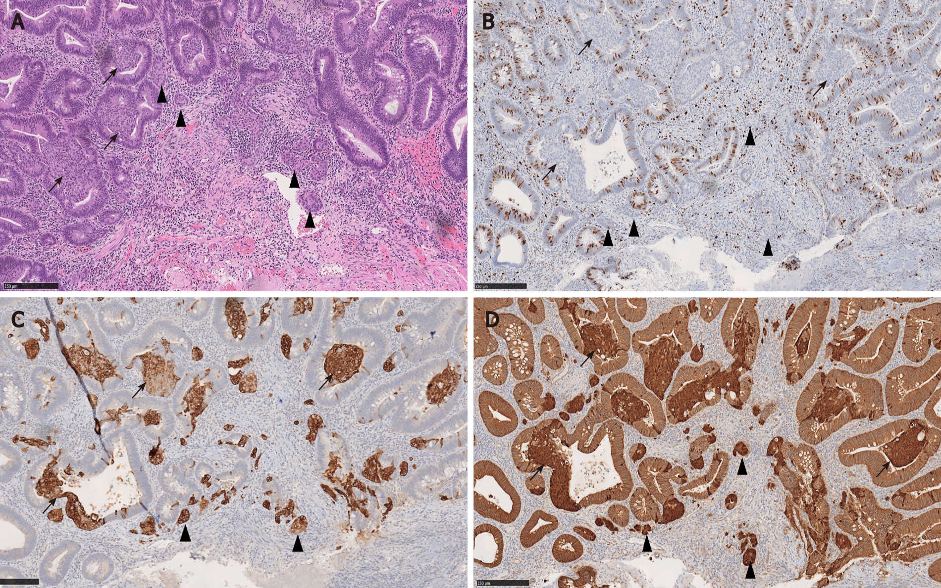
Figure 4 Composite intestinal adenoma-microcarcinoid with associated squamous morules (arrows).
Both microcarcinoid (arrowheads) and squamous morules (arrows) show low proliferative rate and positivity for CK5/6 and beta-catenin (nuclear staining), suggestive of a shared pathogenesis. A: Hematoxylin and eosin, 100 ×; B: Ki 67 immunostain, 100 ×; C: CK5/6 immunostain, 100 ×; D: Beta-catenin immunostain, 100 ×.
Figure 5 Microcarcinoid component of composite intestinal adenoma-microcarcinoid may mimic invasive adenocarcinoma.
A: The microcarcinoid component is found at the base of full-thickness adenomatous glands (Hematoxylin and eosin, 100 ×); B: However, the constituting cells are positive for synaptophysin immunostain (Hematoxylin and eosin, 100 ×).
Figure 6 Rectal neuroendocrine tumor forming a nodule/polyp.
A: The epicenter of the tumor is in the submucosa and the tumor extends to the deep margin (Hematoxylin and eosin, 40 ×); B: High magnification view shows typical trabecular growth pattern (Hematoxylin and eosin, 200 ×).
Figure 7 Appendiceal goblet cell adenocarcinoma.
A: The tumor nests infiltrate and undermine the appendiceal mucosa (Hematoxylin and eosin, 50 ×); B: Bland cytology may mimic well-differentiated neuroendocrine tumor such as seen in the microcarcinoid component of composite intestinal adenoma-microcarcinoid (Hematoxylin and eosin, 200 ×).
Figure 8 Mixed neuroendocrine-non-neuroendocrine neoplasm.
A: Mixed colonic adenocarcinoma (upper right) and large cell neuroendocrine carcinoma (lower left); B: Mixed colonic squamous cell carcinoma and large cell neuroendocrine carcinoma (Inset: high magnification view shows squamous differentiation). Hematoxylin and eosin, 100 ×.
- Citation: Fu ZY, Kmeid M, Aldyab M, Lagana SM, Lee H. Composite intestinal adenoma-microcarcinoid: An update and literature review. World J Gastrointest Endosc 2021; 13(12): 593-606
- URL: https://www.wjgnet.com/1948-5190/full/v13/i12/593.htm
- DOI: https://dx.doi.org/10.4253/wjge.v13.i12.593


