Copyright
©The Author(s) 2021.
World J Gastrointest Endosc. Nov 16, 2021; 13(11): 555-564
Published online Nov 16, 2021. doi: 10.4253/wjge.v13.i11.555
Published online Nov 16, 2021. doi: 10.4253/wjge.v13.i11.555
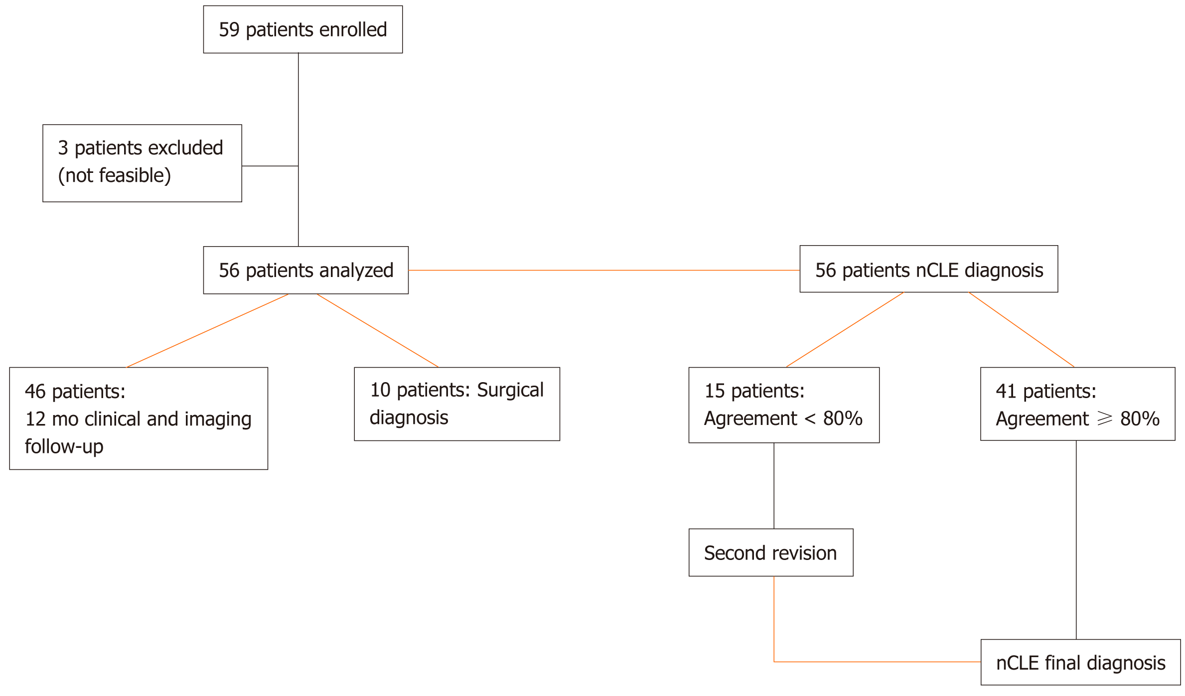
Figure 1 Flow chart.
nCLE: Needle-based confocal endomicroscopy.
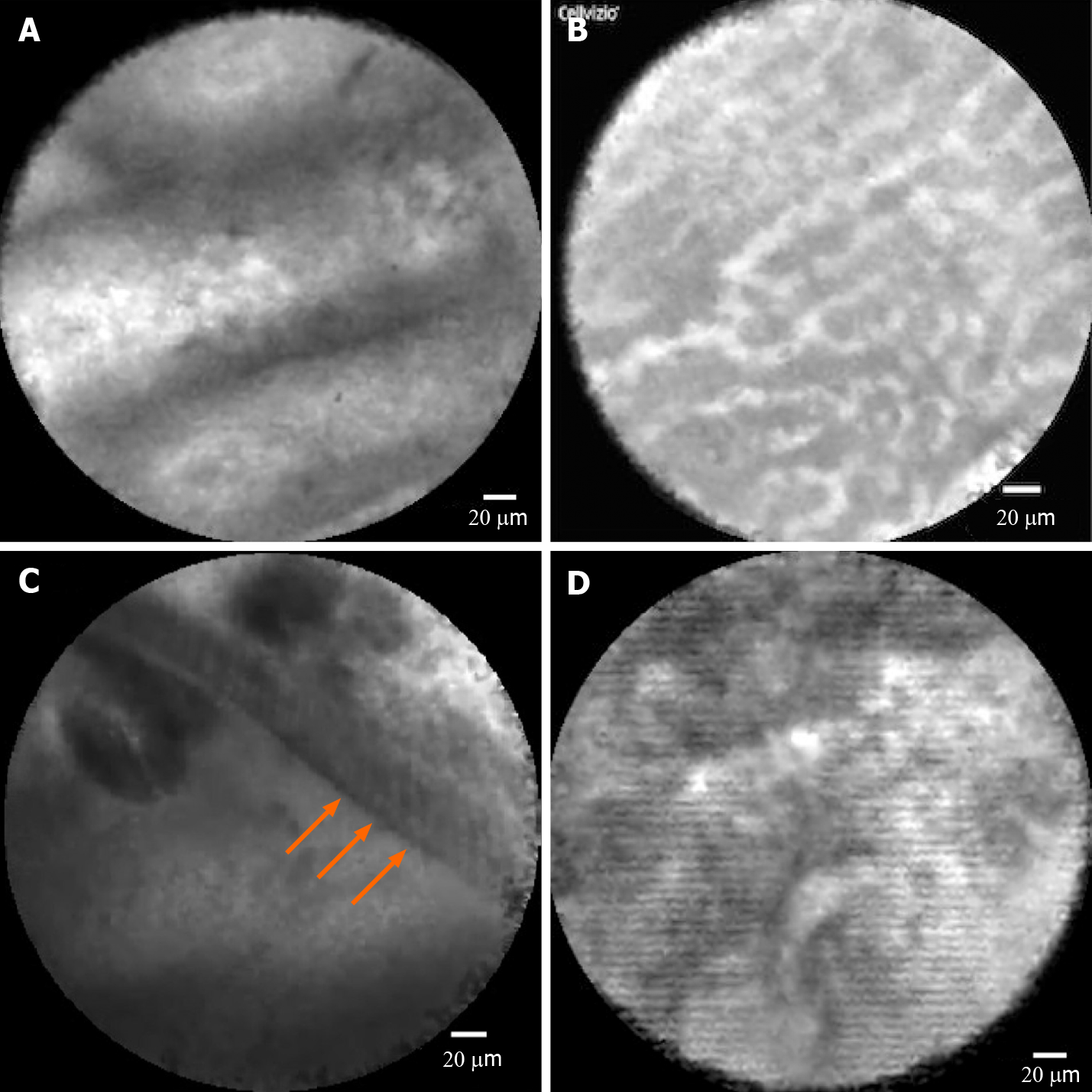
Figure 2 Confocal images of pancreatic cyst subtypes.
A: Intraductal papillary mucinous neoplasm, showing papillary projections; B: Serous cystadenoma, showing superficial vascular network; C: Mucinous cystic neoplasm, in which the epithelial cyst border appears as a gray band delineated by a thin dark line; D: Pseudocyst, showing gray and black particles.
- Citation: Bertani H, Pezzilli R, Pigò F, Bruno M, De Angelis C, Manfredi G, Delconte G, Conigliaro R, Buscarini E. Needle-based confocal endomicroscopy in the discrimination of mucinous from non-mucinous pancreatic cystic lesions. World J Gastrointest Endosc 2021; 13(11): 555-564
- URL: https://www.wjgnet.com/1948-5190/full/v13/i11/555.htm
- DOI: https://dx.doi.org/10.4253/wjge.v13.i11.555


