Copyright
©The Author(s) 2020.
World J Gastrointest Endosc. Sep 16, 2020; 12(9): 297-303
Published online Sep 16, 2020. doi: 10.4253/wjge.v12.i9.297
Published online Sep 16, 2020. doi: 10.4253/wjge.v12.i9.297
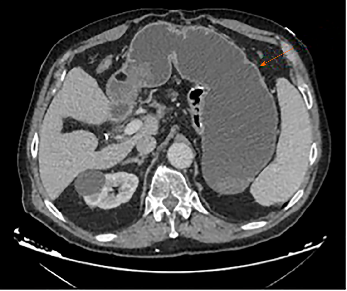
Figure 1 Computerized tomography scan revealing gastric remnant outlet obstruction.
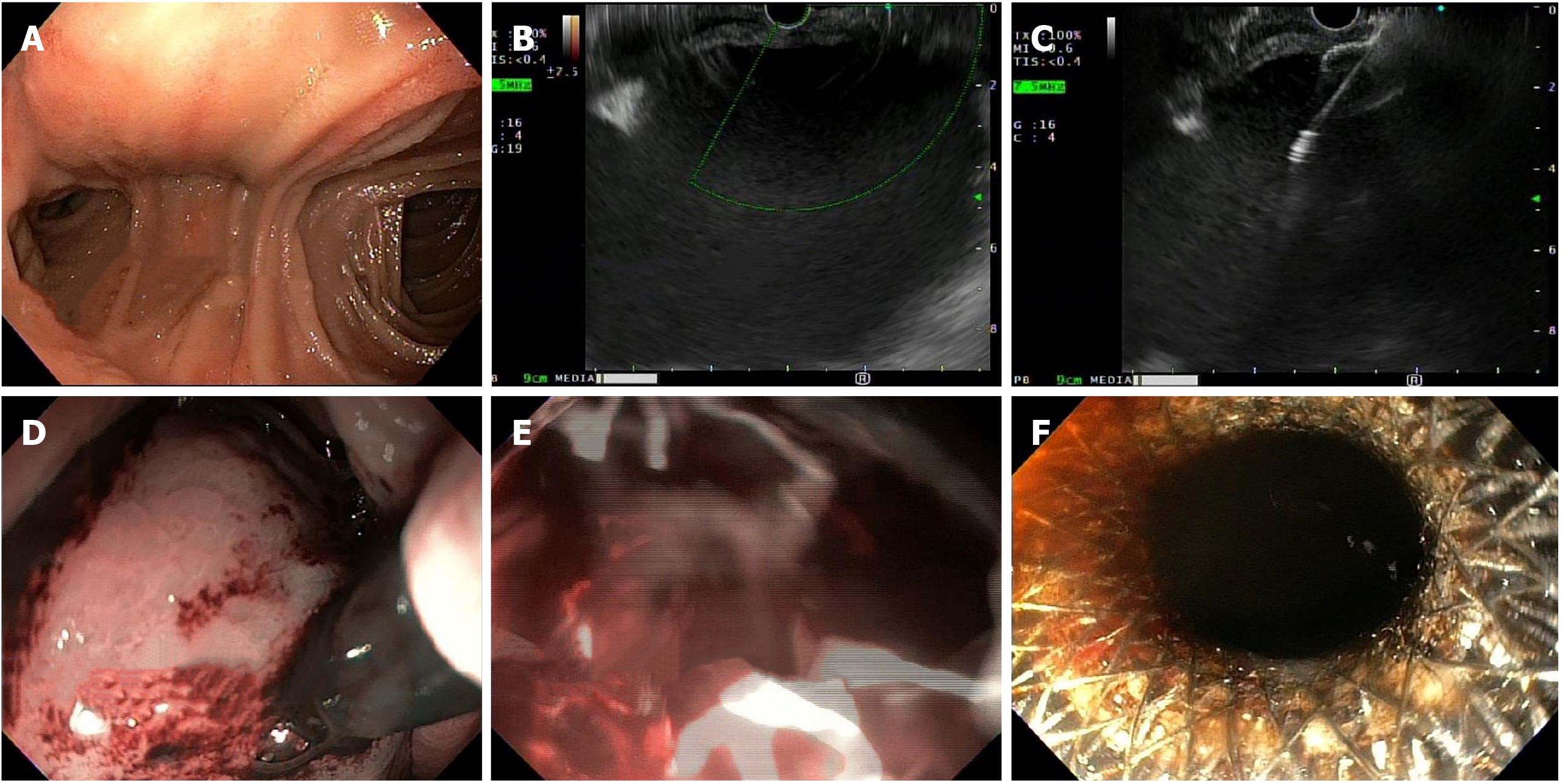
Figure 2 Endoscopy.
A: Healthy-appearing mucosa of Roux-en-Y; B: Gastric distension visualized on Endoscopic ultrasound; C: Introduction of Lumen-Apposing Metal Stent; D: Endoscopic view of Lumen-Apposing Metal Stent placement; E: Immediate decompression with flow of blood through stent; and F: Successful endoscopic ultrasound guided gastro-gastrostomy using Lumen-Apposing Metal Stent.
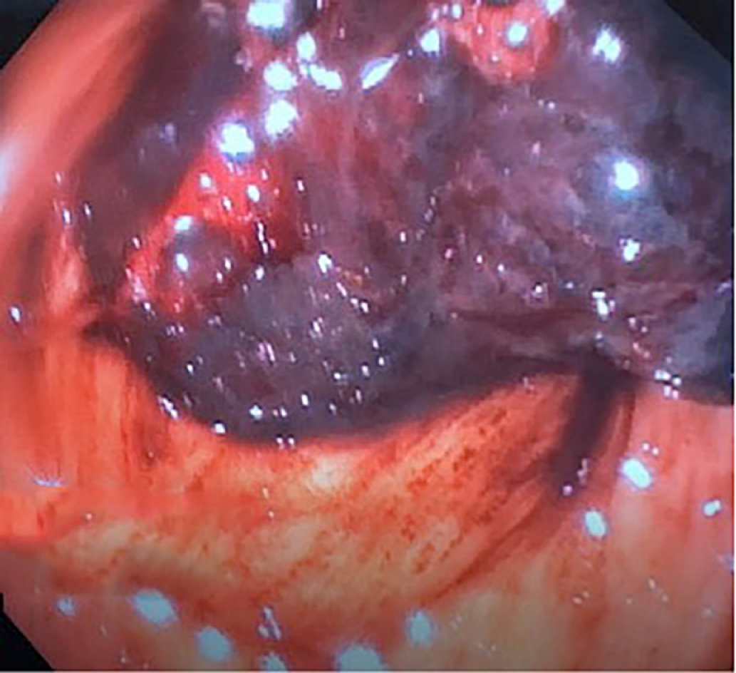
Figure 3 A large clot in gastric antrum of the excluded stomach.
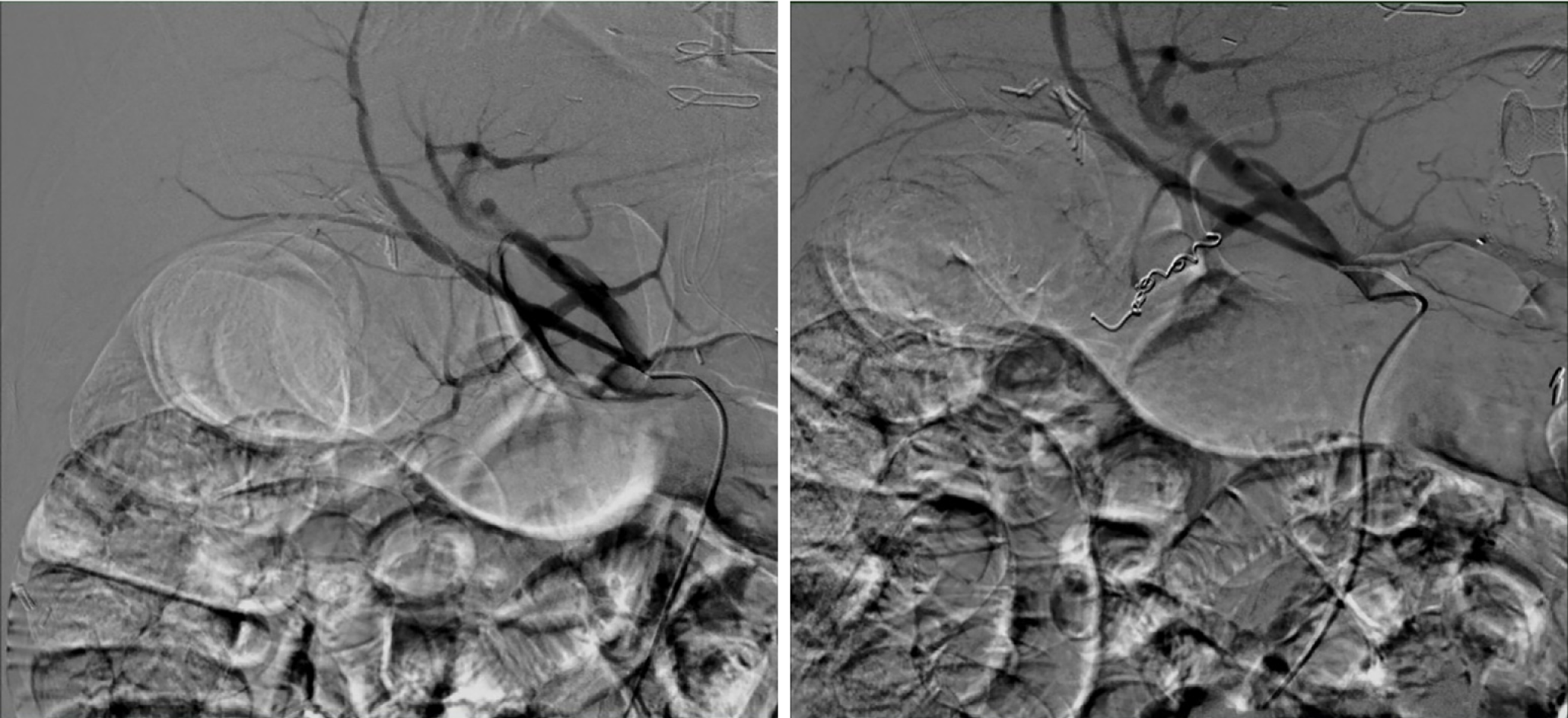
Figure 4 Angiography and embolization of gastroduodenal artery.
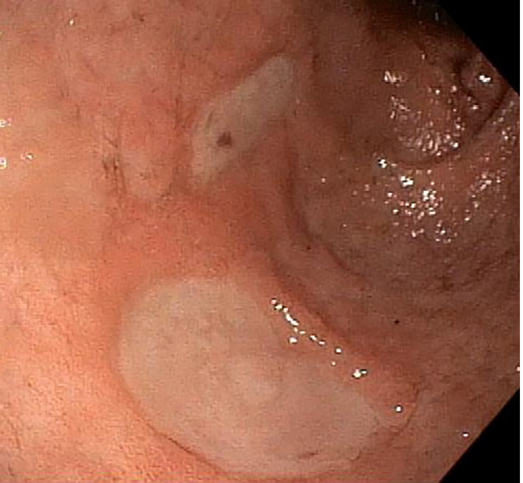
Figure 5 Healing gastric ulcers on follow up endoscopy.
- Citation: Zarrin A, Sorathia S, Choksi V, Kaplan SR, Kasmin F. Endoscopic approach to gastric remnant outlet obstruction after gastric bypass: A case report. World J Gastrointest Endosc 2020; 12(9): 297-303
- URL: https://www.wjgnet.com/1948-5190/full/v12/i9/297.htm
- DOI: https://dx.doi.org/10.4253/wjge.v12.i9.297


