Copyright
©The Author(s) 2020.
World J Gastrointest Endosc. May 16, 2020; 12(5): 149-158
Published online May 16, 2020. doi: 10.4253/wjge.v12.i5.149
Published online May 16, 2020. doi: 10.4253/wjge.v12.i5.149
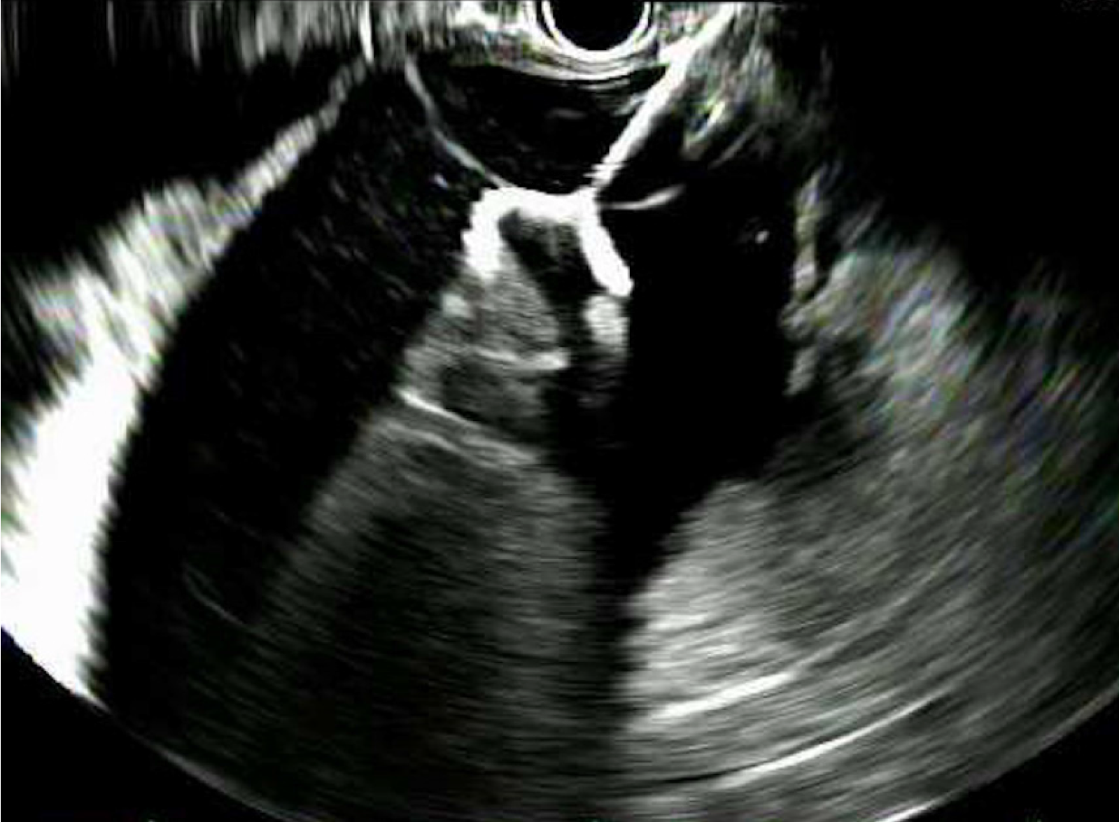
Figure 1 Sonographic guided deployment of distal lumen apposing metal stent flange.
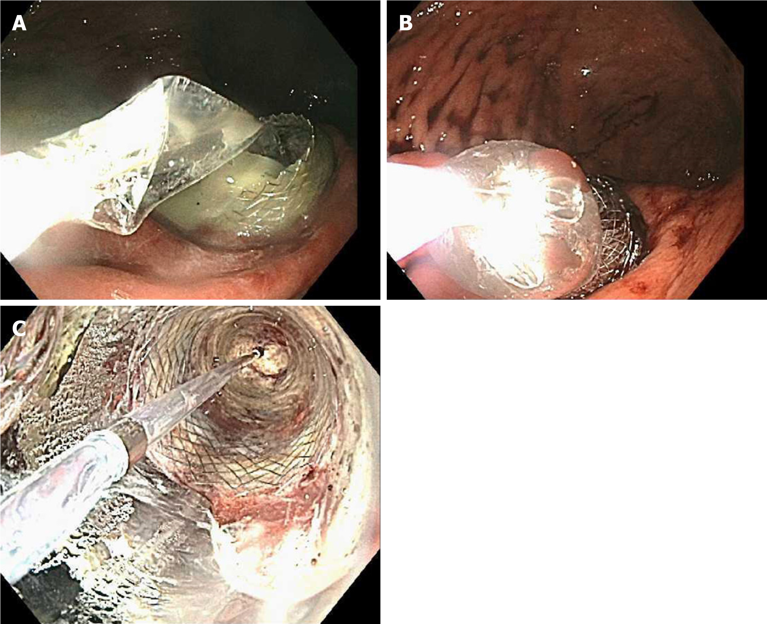
Figure 2 Endoscopy results.
A-C: Dilation of the lumen apposing metal stent lumen with a through-the-scope balloon.
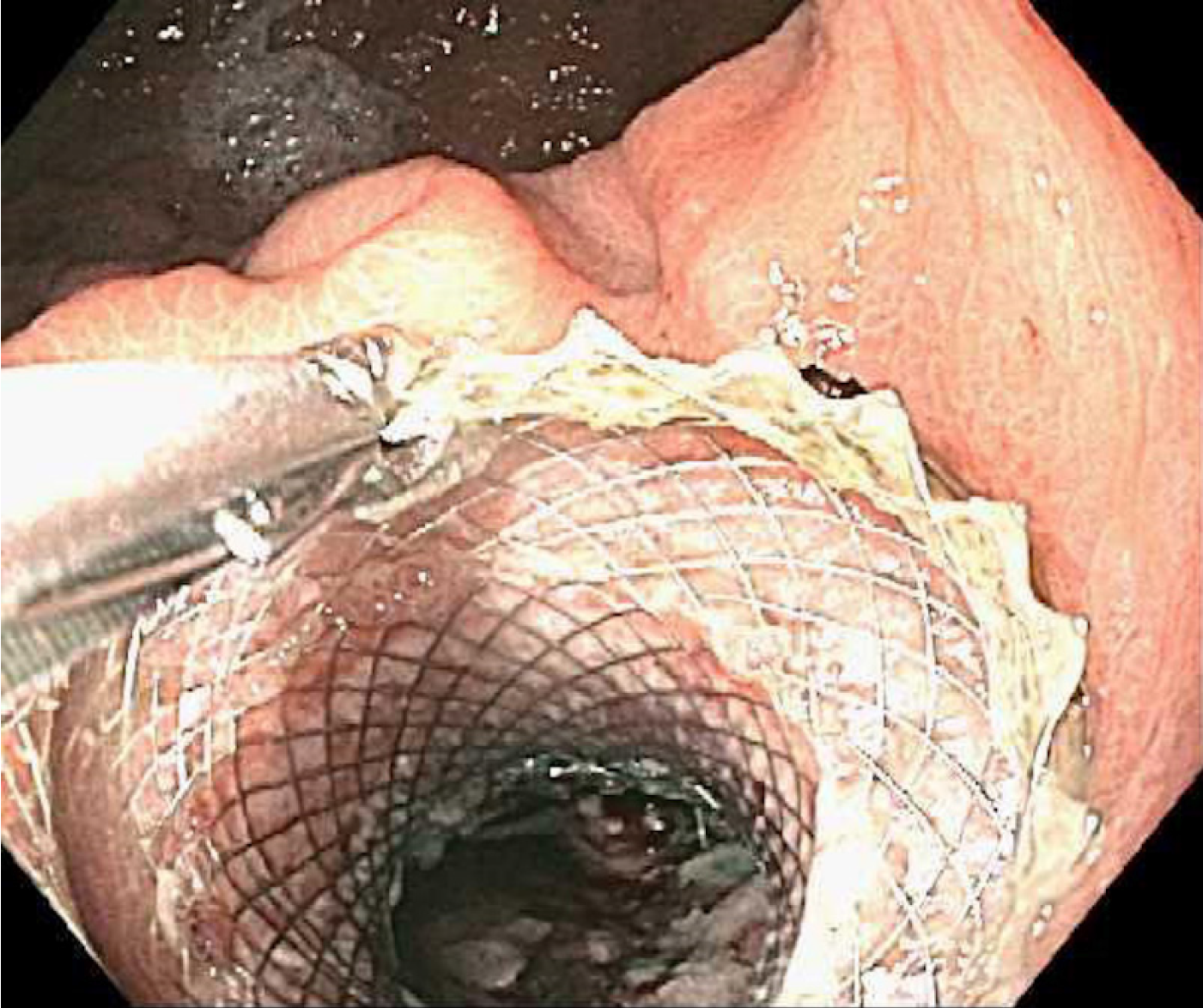
Figure 3 Endoscopy results.
A: Debridement of necrosum with a metal snare; B: Clean walled-off necrosis cavity after debridement.
Figure 4 Removal of lumen apposing metal stent.
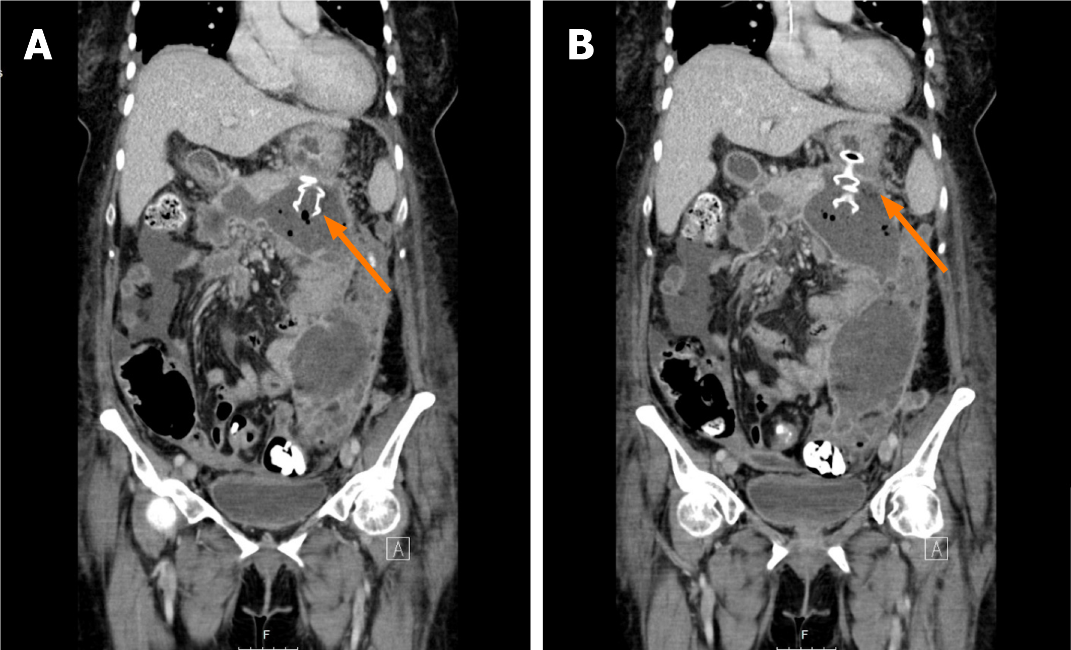
Figure 5 Computed tomography of the abdomen.
A: Lumen apposing metal stent mis-deployed inside the walled-off necrosis cavity; B: Second lumen apposing metal stent successfully placed through the original puncture site.
- Citation: Mendoza Ladd A, Bashashati M, Contreras A, Umeanaeto O, Robles A. Endoscopic pancreatic necrosectomy in the United States-Mexico border: A cross sectional study. World J Gastrointest Endosc 2020; 12(5): 149-158
- URL: https://www.wjgnet.com/1948-5190/full/v12/i5/149.htm
- DOI: https://dx.doi.org/10.4253/wjge.v12.i5.149


