Copyright
©The Author(s) 2020.
World J Gastrointest Endosc. Jan 16, 2020; 12(1): 42-48
Published online Jan 16, 2020. doi: 10.4253/wjge.v12.i1.42
Published online Jan 16, 2020. doi: 10.4253/wjge.v12.i1.42
Figure 1 Gastrografin swallow showing esophagogastric leakage.
Figure 2 Gastroscopy results.
A: Dehiscence of the anastomosis with 30% of the circumference; B: Anastomotic leak in the suture line; C: Endoscopic vacuum assisted closure (E-VAC) introduced into the mediastinal cavity; D: Mediastinal cavity reduced to a small and shallow like an esophageal diverticulum; E: E-VAC introduced into the lumen of the gastrointestinal tract close to the anastomotic leak; F: Follow-up gastroscopy: anastomotic stricture with shallow diverticulum.
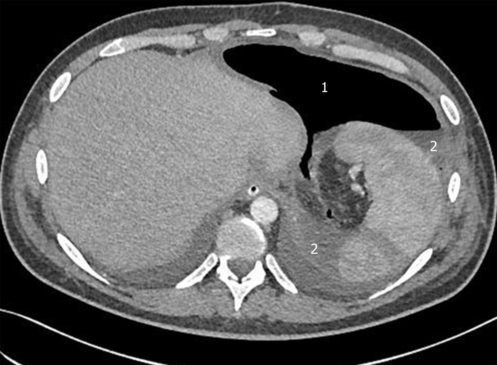
Figure 3 Computed tomography on the third day after relaparotomy.
A large amount of gas (1) and liquid (2) as a result of leakage anastomosis.
Figure 4 Endoscopic vacuum assisted closure prepared before the introduction.
- Citation: Cwaliński J, Hermann J, Kasprzyk M, Banasiewicz T. Endoscopic vacuum assisted closure of esophagogastric anastomosis dehiscence: A case report. World J Gastrointest Endosc 2020; 12(1): 42-48
- URL: https://www.wjgnet.com/1948-5190/full/v12/i1/42.htm
- DOI: https://dx.doi.org/10.4253/wjge.v12.i1.42


