Copyright
©The Author(s) 2019.
World J Gastrointest Endosc. May 16, 2019; 11(5): 329-344
Published online May 16, 2019. doi: 10.4253/wjge.v11.i5.329
Published online May 16, 2019. doi: 10.4253/wjge.v11.i5.329
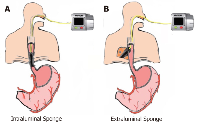
Figure 1 Sponge placement.
A: Intraluminal endoscopic vacuum therapy (EVT); B: Intracavitary EVT.
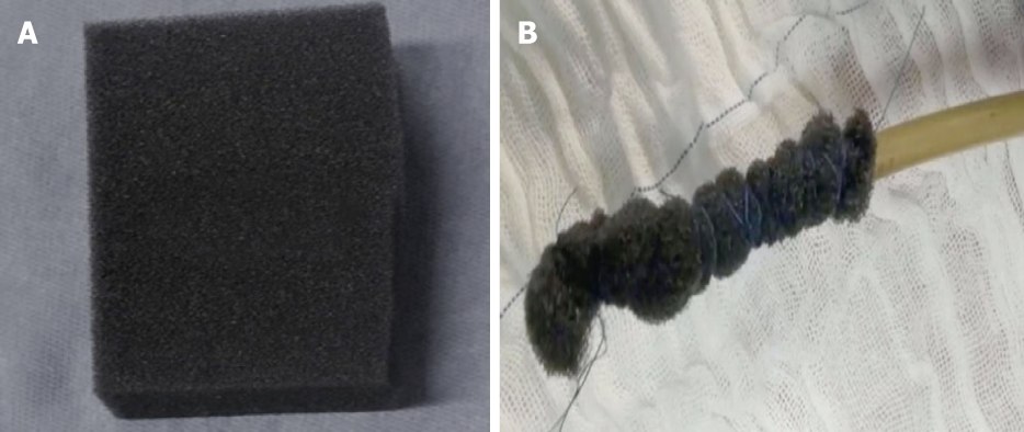
Figure 2 Open-pore polyurethane sponge drain.
A: Open-pore polyurethane sponge; B: Open-pore polyurethane sponge drain for endoscopic vacuum therapy.
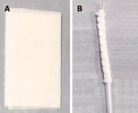
Figure 3 Open-pore film drain.
A: Open-pore film; B: Open-pore film drain for endoscopic vacuum therapy.
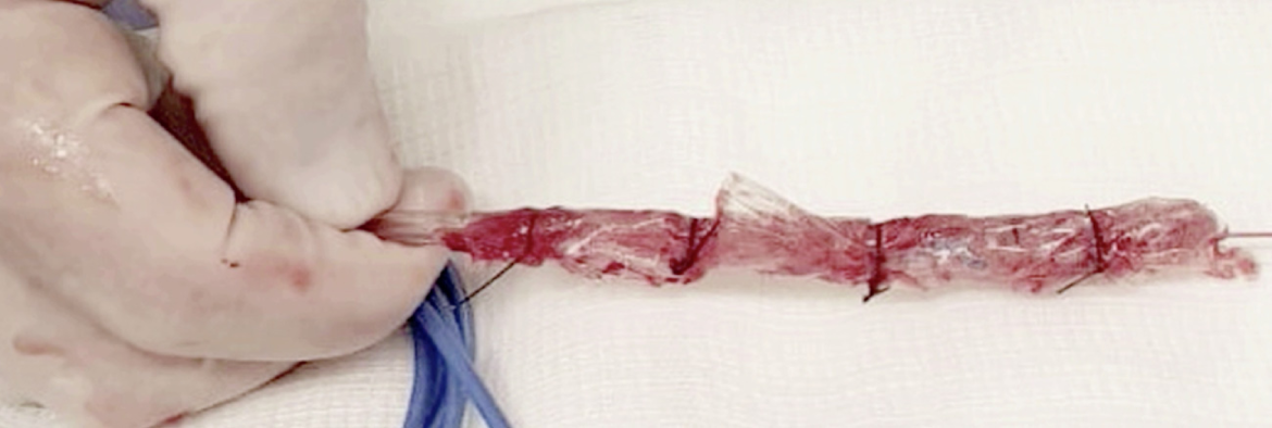
Figure 4 A low cost modified endoscopic vacuum therapy drain system made with a gauze coated with perforated sterile plastic.
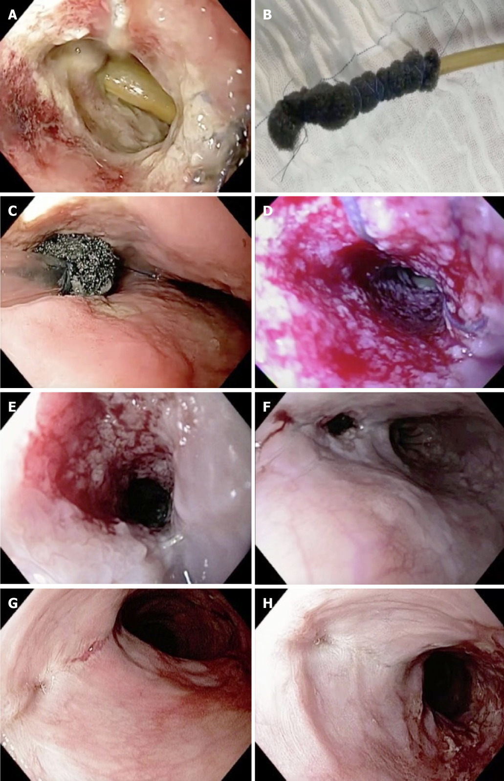
Figure 5 Endoscopic vacuum therapy in the management of an esophageal defect.
A: Complete dehiscence of the esophageal leak and the mediastinal drainage; B: Open-pore polyurethane foam drain; C: Intracavitary sponge placement; D: Granulation tissue after second sponge exchange; E: Granulation tissue after fourth sponge exchange; F: Reduction of the defect size after seven sponge exchanges; G: Complete closure after nine sponge exchanges; H: Scar after esophageal closure with endoscopic vacuum therapy.
- Citation: de Moura DTH, de Moura BFBH, Manfredi MA, Hathorn KE, Bazarbashi AN, Ribeiro IB, de Moura EGH, Thompson CC. Role of endoscopic vacuum therapy in the management of gastrointestinal transmural defects. World J Gastrointest Endosc 2019; 11(5): 329-344
- URL: https://www.wjgnet.com/1948-5190/full/v11/i5/329.htm
- DOI: https://dx.doi.org/10.4253/wjge.v11.i5.329


