Copyright
©The Author(s) 2019.
World J Gastrointest Endosc. Apr 16, 2019; 11(4): 281-291
Published online Apr 16, 2019. doi: 10.4253/wjge.v11.i4.281
Published online Apr 16, 2019. doi: 10.4253/wjge.v11.i4.281
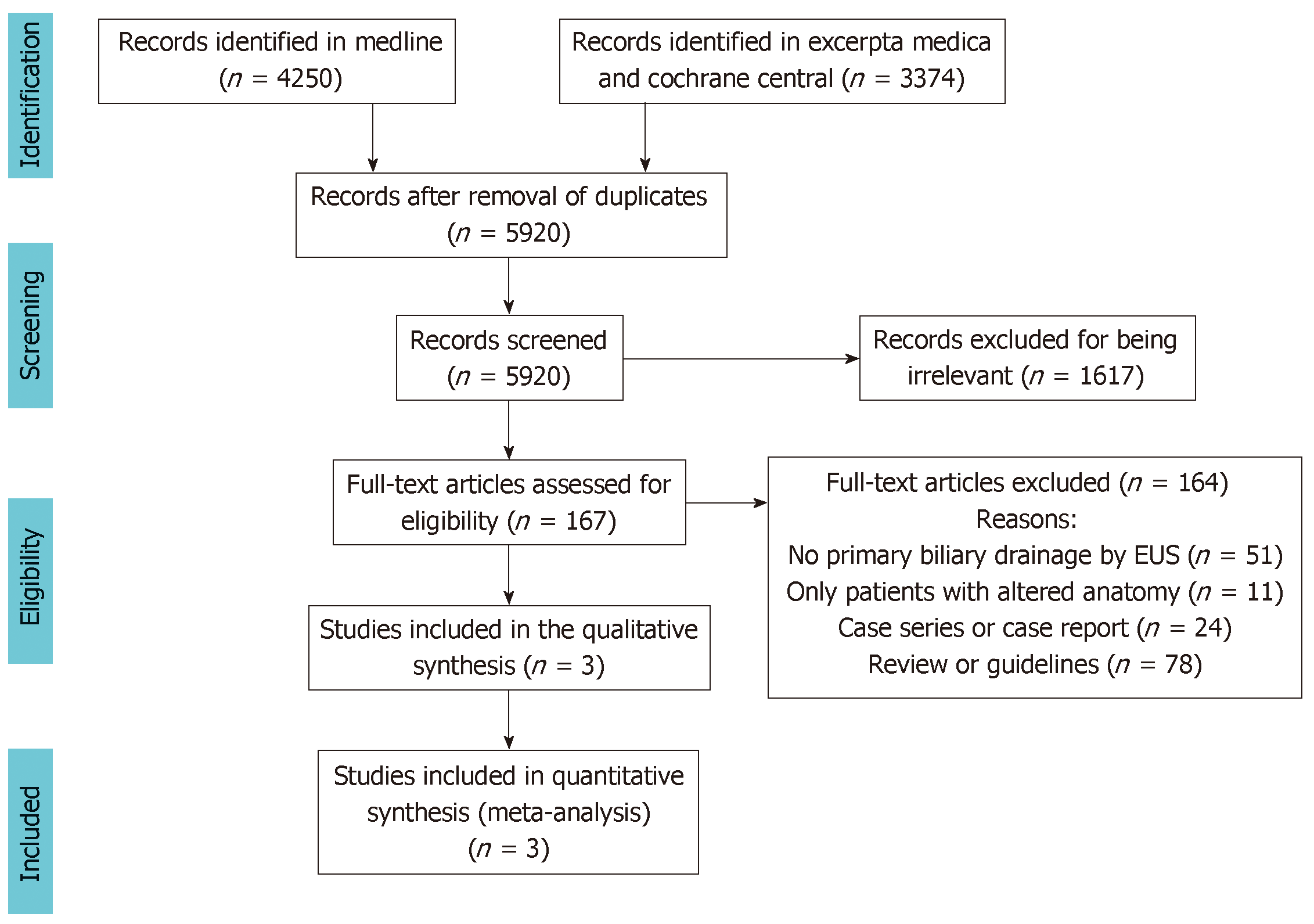
Figure 1 Flow chart of study selection.
Cochrane CENTRAL: Cochrane Central Register of Controlled Trials; EUS: Endoscopic ultrasound.
Figure 2 Forest plot of technical success.
M-H: Mantel-Haenszel test; EUS: Endoscopic ultrasound; ERCP: Endoscopic retrograde cholangiopancreatography.
Figure 3 Forest plot of clinical success.
M-H: Mantel-Haenszel test; EUS: Endoscopic ultrasound; ERCP: Endoscopic retrograde cholangiopancreatography.
Figure 4 Forest plot of procedure duration in minutes.
IV: Inverse variance test; EUS: Endoscopic ultrasound; ERCP: Endoscopic retrograde cholangiopancreatography.
Figure 5 Forest plot of adverse events.
M-H: Mantel-Haenszel test; EUS: Endoscopic ultrasound; ERCP: Endoscopic retrograde cholangiopancreatography.
Figure 6 Forest plot of stent patency.
IV: Inverse variance test; EUS: Endoscopic ultrasound; ERCP: Endoscopic retrograde cholangiopancreatography.
Figure 7 Forest plot of stent dysfunction requiring intervention.
M-H: Mantel-Haenszel test; EUS: Endoscopic ultrasound; ERCP: Endoscopic retrograde cholangiopancreatography.
- Citation: Logiudice FP, Bernardo WM, Galetti F, Sagae VM, Matsubayashi CO, Madruga Neto AC, Brunaldi VO, de Moura DTH, Franzini T, Cheng S, Matuguma SE, de Moura EGH. Endoscopic ultrasound-guided vs endoscopic retrograde cholangiopancreatography biliary drainage for obstructed distal malignant biliary strictures: A systematic review and meta-analysis. World J Gastrointest Endosc 2019; 11(4): 281-291
- URL: https://www.wjgnet.com/1948-5190/full/v11/i4/281.htm
- DOI: https://dx.doi.org/10.4253/wjge.v11.i4.281


