Copyright
©The Author(s) 2019.
World J Gastrointest Endosc. Mar 16, 2019; 11(3): 256-261
Published online Mar 16, 2019. doi: 10.4253/wjge.v11.i3.256
Published online Mar 16, 2019. doi: 10.4253/wjge.v11.i3.256
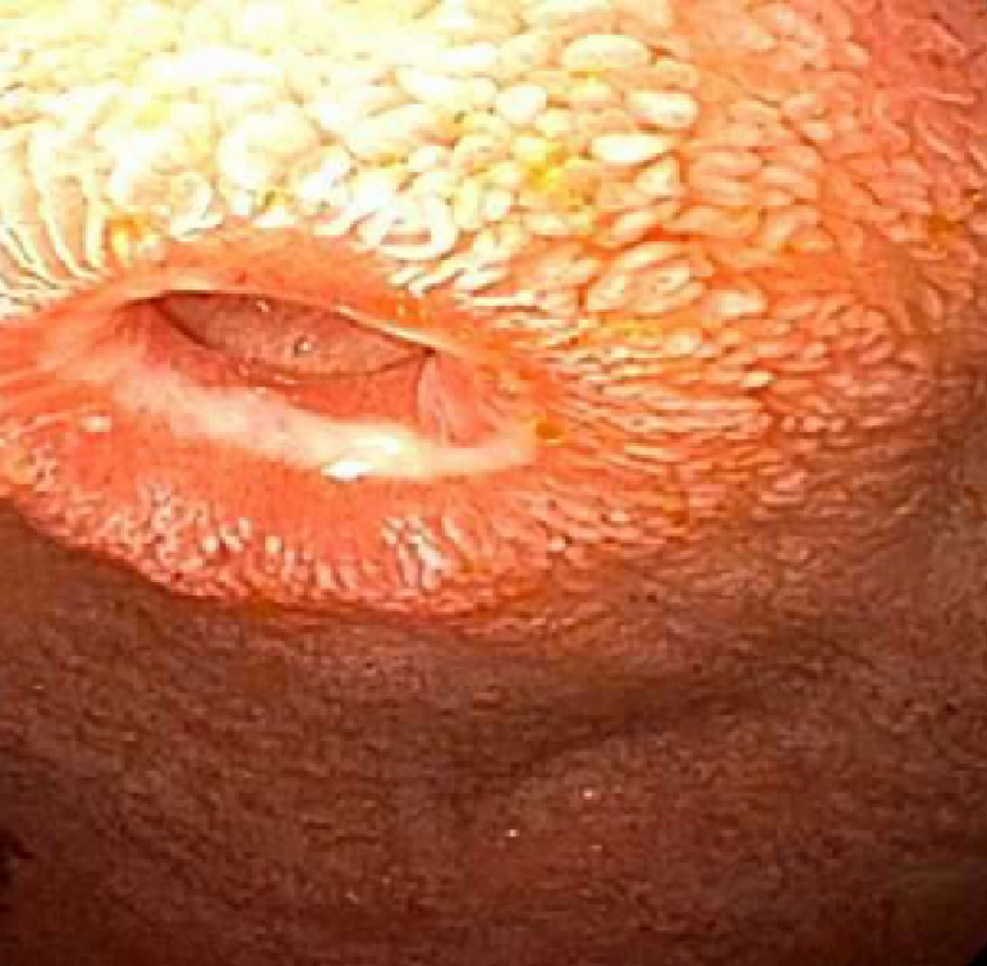
Figure 1 Endoscopic view of the duodenal bulb with the stricture.
Figure 2 Upper gastrointestinal series revealing the extent of the duodenal stricture.
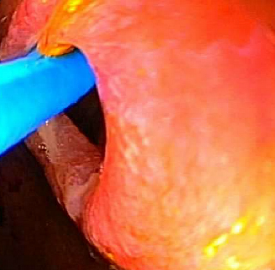
Figure 3 Endoscopic view showing the choledochoscope advanced out from the therapeutic gastroscope traverse across the stricture.
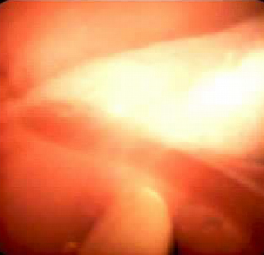
Figure 4 Endoscopic view from the choledochoscope with direct visualization of the stenosis.
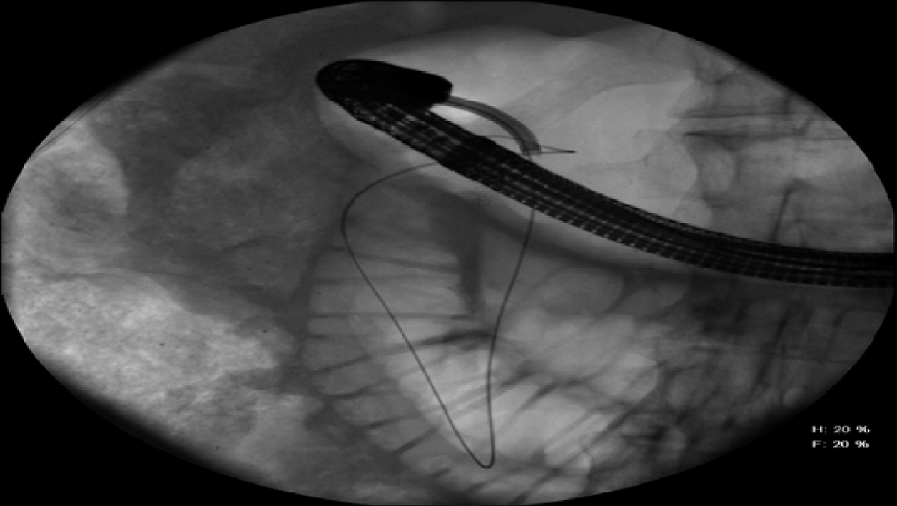
Figure 5 Radiographic view of the choledochoscope (advanced through the therapeutic gastroscope) crossing the duodenal stenosis and allowing passage of the guidewire.
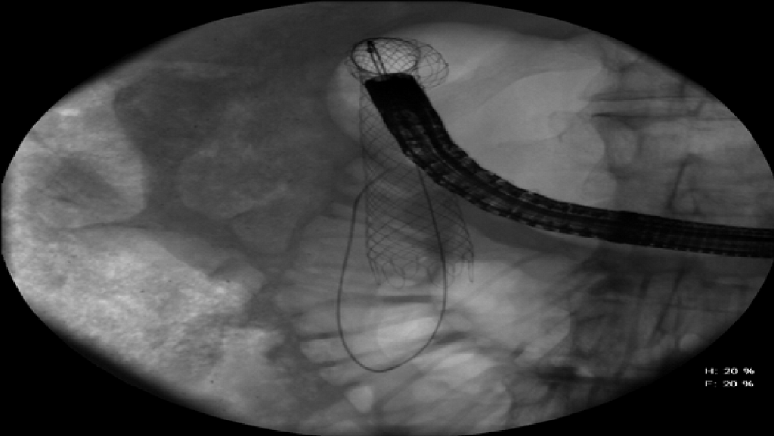
Figure 6 Radiographic view of the guidewire-directed stent placement crossing the duodenal stenosis.
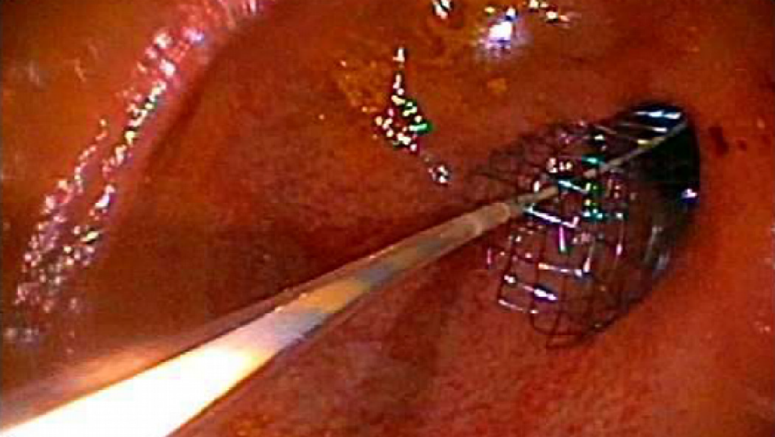
Figure 7 Endoscopic view of the stent deployment.
- Citation: Cho RSE, Magulick J, Madden S, Burdick JS. Choledochoscope with stent placement for treatment of benign duodenal strictures: A case report. World J Gastrointest Endosc 2019; 11(3): 256-261
- URL: https://www.wjgnet.com/1948-5190/full/v11/i3/256.htm
- DOI: https://dx.doi.org/10.4253/wjge.v11.i3.256


