Copyright
©The Author(s) 2019.
World J Gastrointest Endosc. Mar 16, 2019; 11(3): 193-208
Published online Mar 16, 2019. doi: 10.4253/wjge.v11.i3.193
Published online Mar 16, 2019. doi: 10.4253/wjge.v11.i3.193
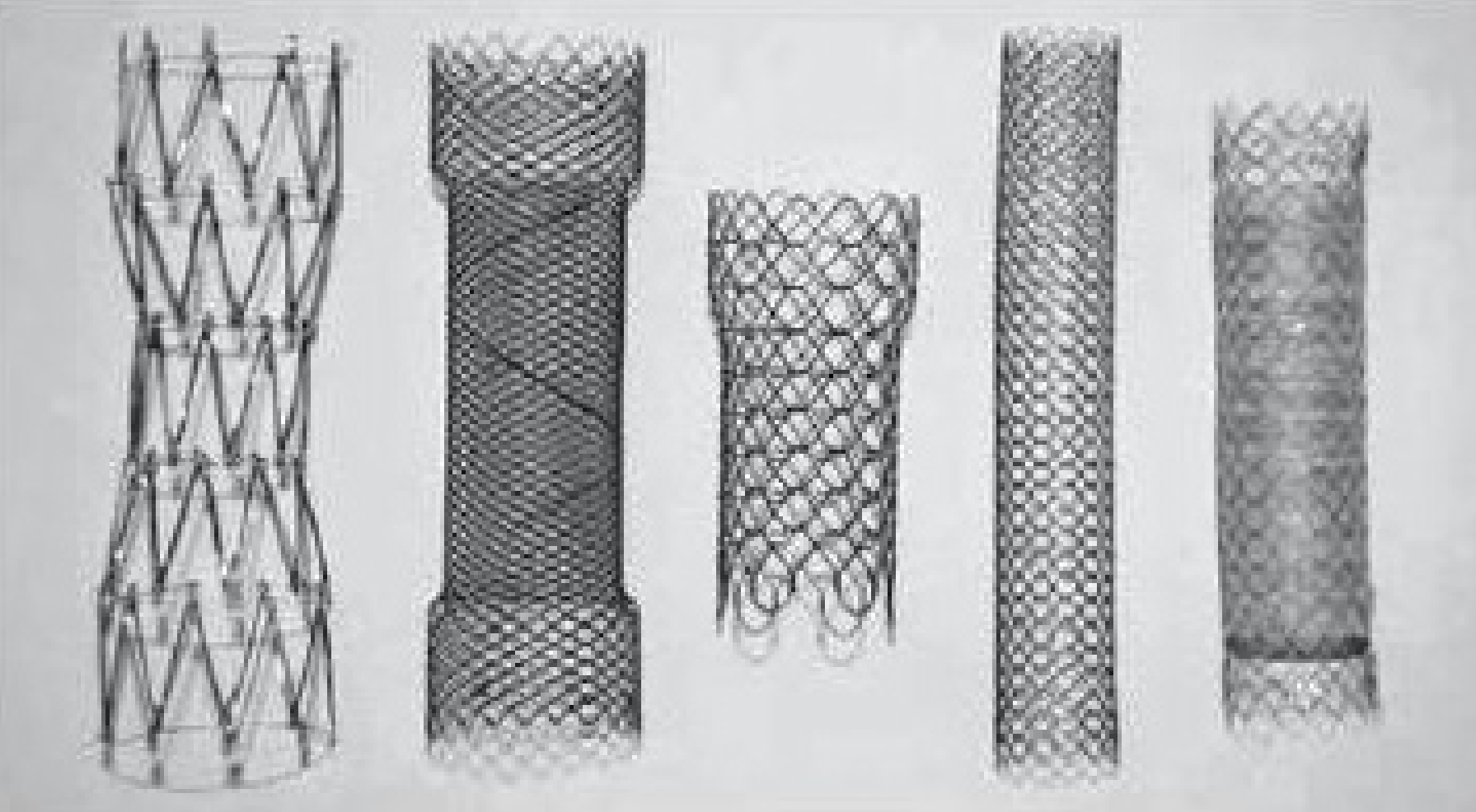
Figure 1 Stent models, left to right: Colonic Z (Cook), Evolution colonic (Cook), Wallflex (Boston), D-type colonic not covered (Taewoog), type colonic covered (Taewoong).
All FDA approved stents.
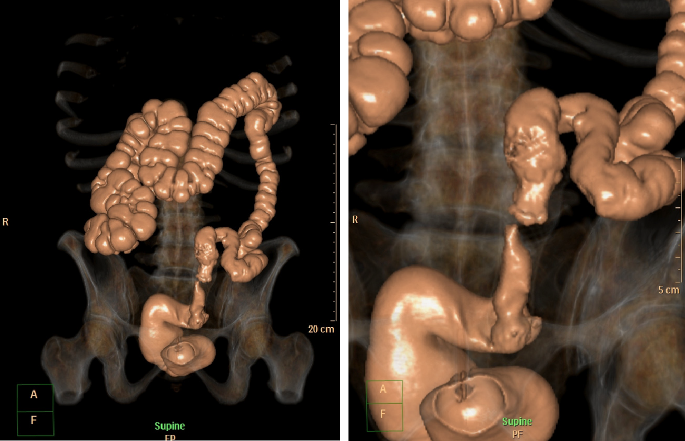
Figure 2 Virtual Colonoscopy showing a malignant stricture in the distal left colon.
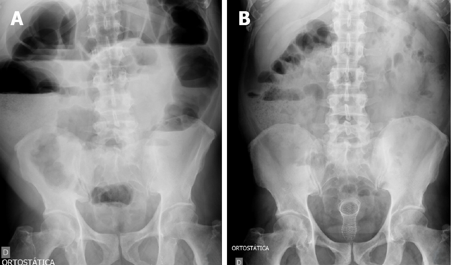
Figure 3 Radiography images.
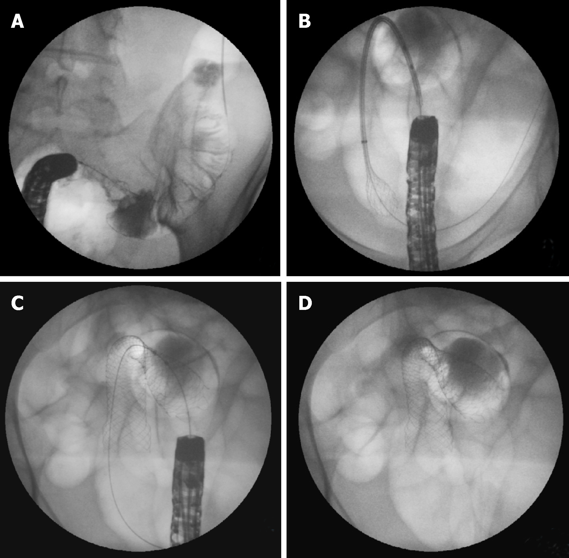
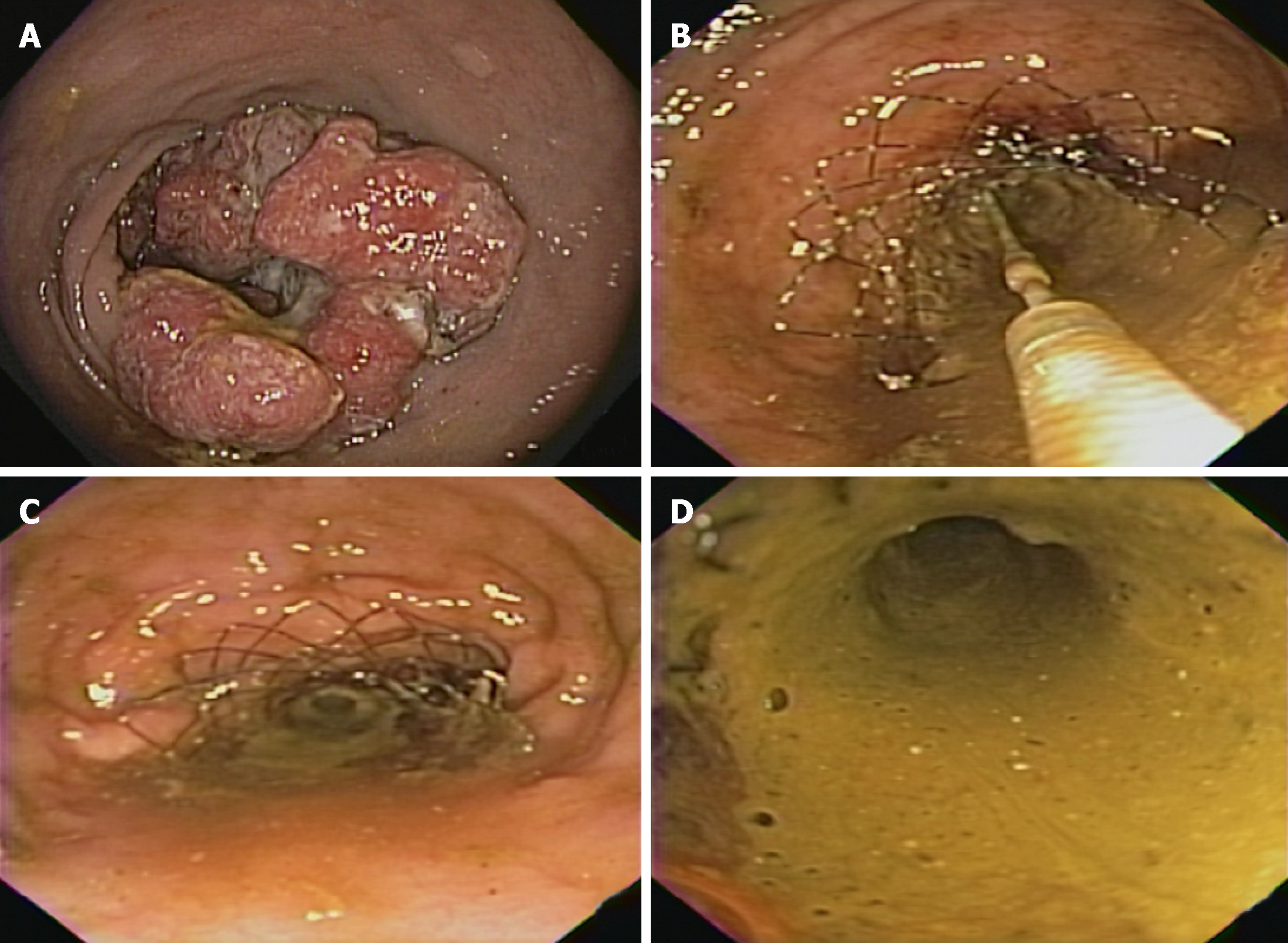
A: Radiography showing signs of a distal obstruction; B: Radiography of the same patient showing improvement after stenting and decompression. Figure 4 Stents procedure. A: Guide-wire placement through the stricture after contrast study; B: Stent deploying through the scope; C: Stent placement showing the stricture in the middle of the stent; D: Final stent position.
Figure 4 Stents procedure.
A: Guide-wire placement through the stricture after contrast study; B: Stent deploying through the scope; C: Stent placement showing the stricture in the middle of the stent; D: Final stent position.
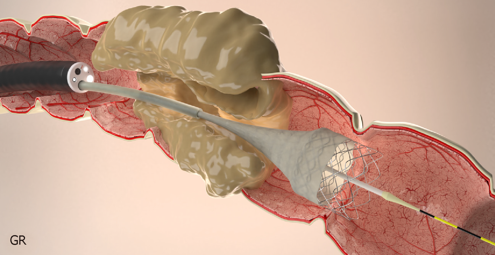
Figure 5 Stent placement by Through the scope technique.
Figure 6 The stent is passed over the guidewire to the proximal margin of the tumor and then implanted under fluoroscopic guidance and endoscopic visualization of the distal portion of the stent.
A: Malignant lesion causing colonic stenosis; B: Stent deployment; C: Stent immediately after deployment; D: Fecal contents coming through the stent after decompression.
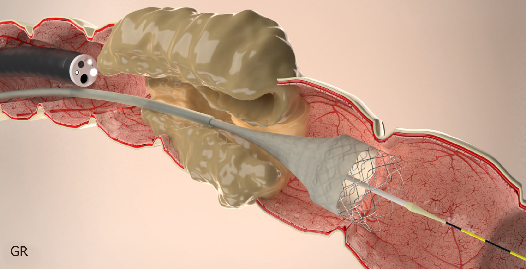
Figure 7 Stent placement by Over-the-wire technique.
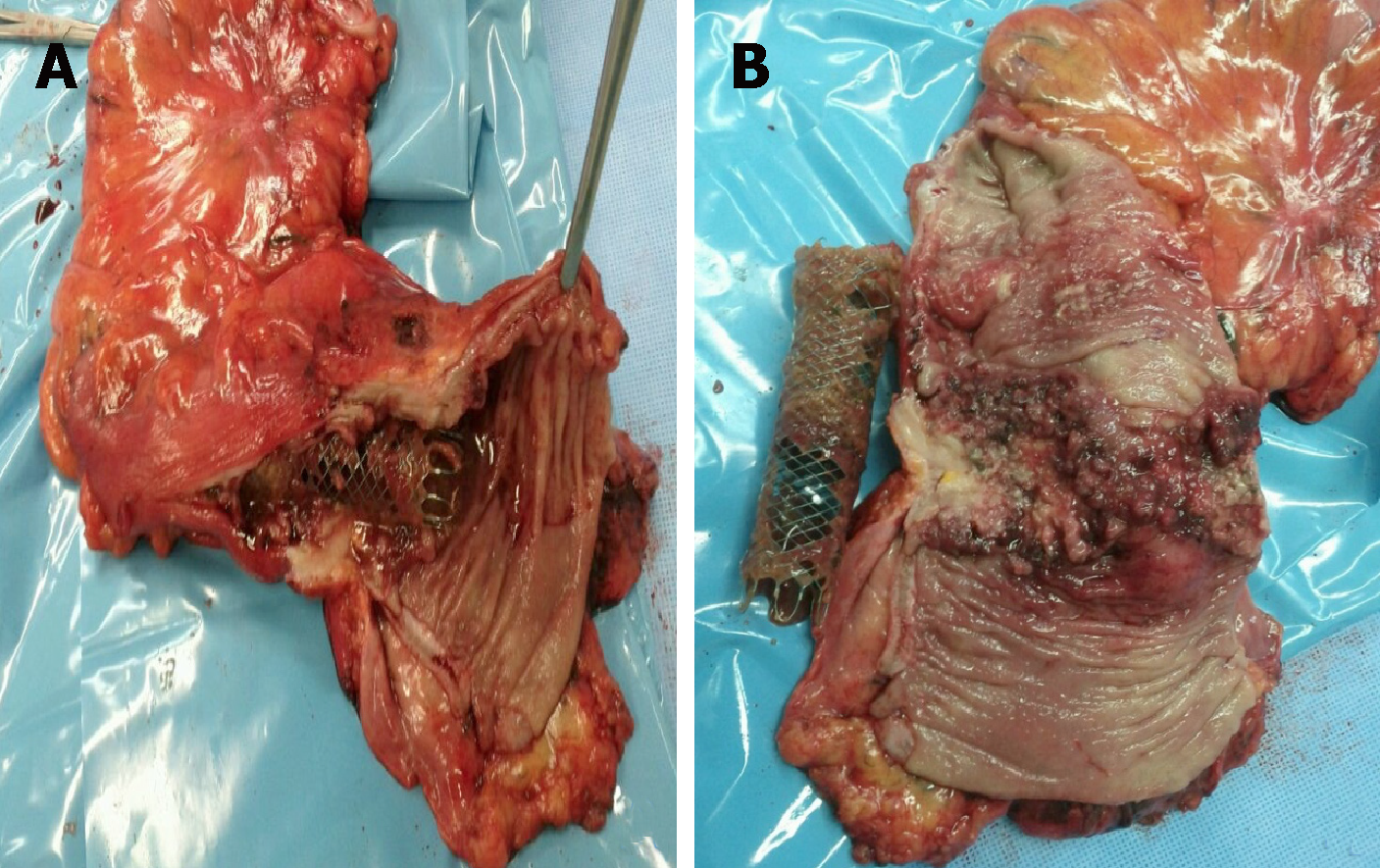
Figure 8 Specimen and stent.
A: Specimen after surgery showing the stent crossing the lesion; B: Image of the stent and the malignant lesion after resection.
- Citation: Ribeiro IB, de Moura DTH, Thompson CC, de Moura EGH. Acute abdominal obstruction: Colon stent or emergency surgery? An evidence-based review. World J Gastrointest Endosc 2019; 11(3): 193-208
- URL: https://www.wjgnet.com/1948-5190/full/v11/i3/193.htm
- DOI: https://dx.doi.org/10.4253/wjge.v11.i3.193


