Copyright
©The Author(s) 2019.
World J Gastrointest Endosc. Nov 16, 2019; 11(11): 541-547
Published online Nov 16, 2019. doi: 10.4253/wjge.v11.i11.541
Published online Nov 16, 2019. doi: 10.4253/wjge.v11.i11.541
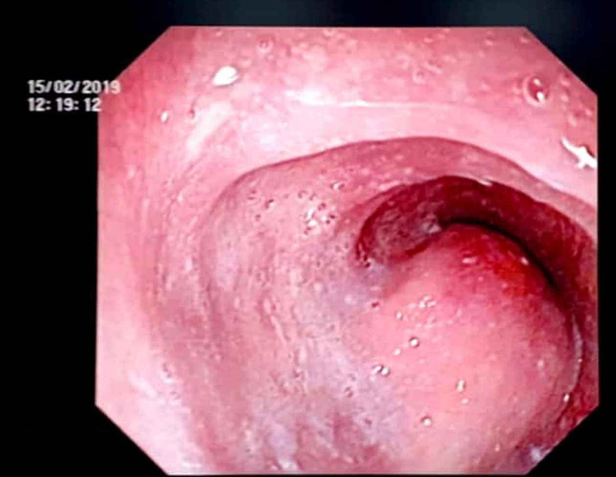
Figure 1 Upper gastrointestinal endoscopy.
A large, hemispherical lesion measuring about 4 cm × 5 cm in size, with a normal-appearing overlying mucosa extending from 30-34 cm from the incisors.
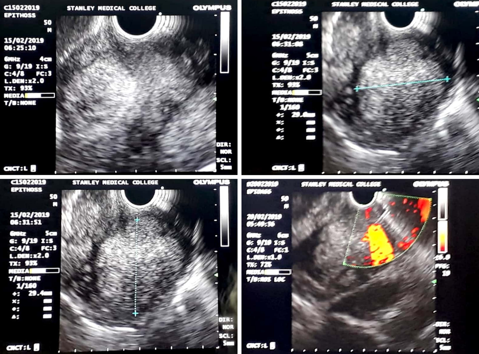
Figure 2 Endoscopic ultrasound images.
Endoscopic ultrasound showing a hyperechoic mass lesion measuring 4 cm × 5 cm arising from the third layer of the oesophagus.
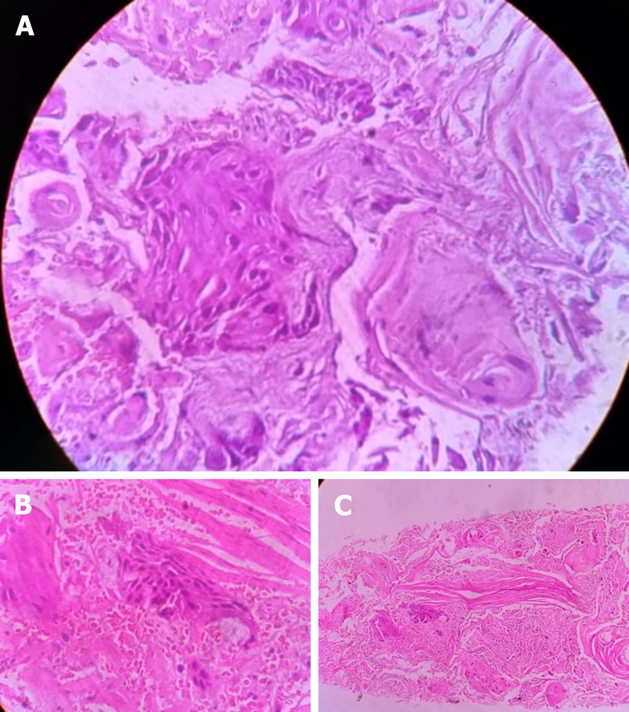
Figure 3 Photomicrography of oesophageal mucosal.
A-C: Photomicrography of oesophageal mucosa showing round-to-polygonal neoplastic cells with moderate amounts of eosinophilic cytoplasm, with moderate nuclear atypia and abundant keratin pearl formation. This is consistent with well-differentiated esophageal squamous cell carcinoma (Hematoxylin-eosin staining).
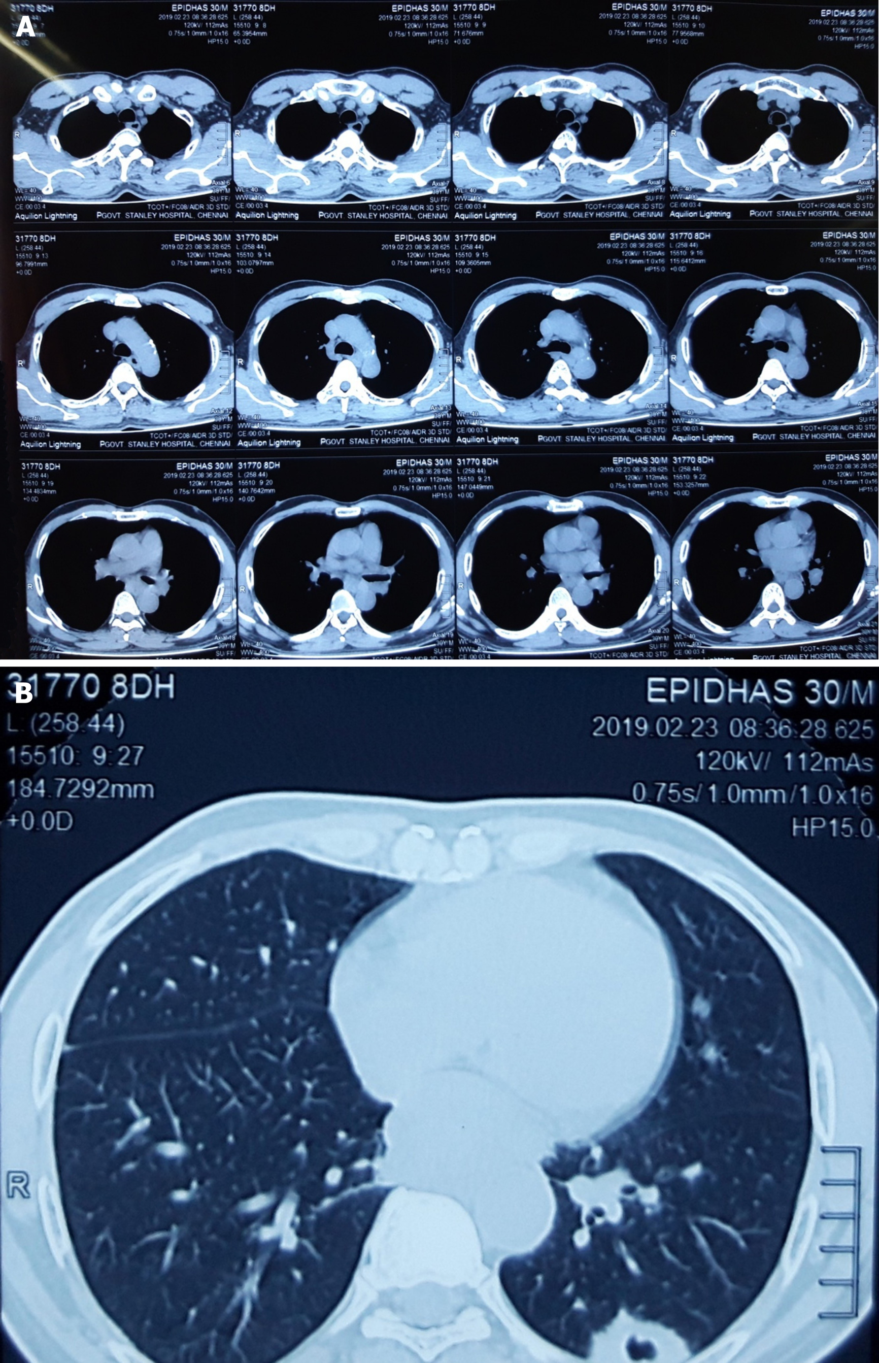
Figure 4 Computed tomography images.
A and B: Computed tomography chest with oral and IV contrast, showing a cavitatory metastatic nodule measuring 26 mm × 18 mm × 33 mm in the posterior basal segment of the left lower lobe.
- Citation: Shanmugam RM, Shanmugam C, Murugesan M, Kalyansundaram M, Gopalsamy S, Ranjan A. Oesophageal carcinoma mimicking a submucosal lesion: A case report. World J Gastrointest Endosc 2019; 11(11): 541-547
- URL: https://www.wjgnet.com/1948-5190/full/v11/i11/541.htm
- DOI: https://dx.doi.org/10.4253/wjge.v11.i11.541


