Copyright
©The Author(s) 2019.
World J Gastrointest Endosc. Nov 16, 2019; 11(11): 531-540
Published online Nov 16, 2019. doi: 10.4253/wjge.v11.i11.531
Published online Nov 16, 2019. doi: 10.4253/wjge.v11.i11.531
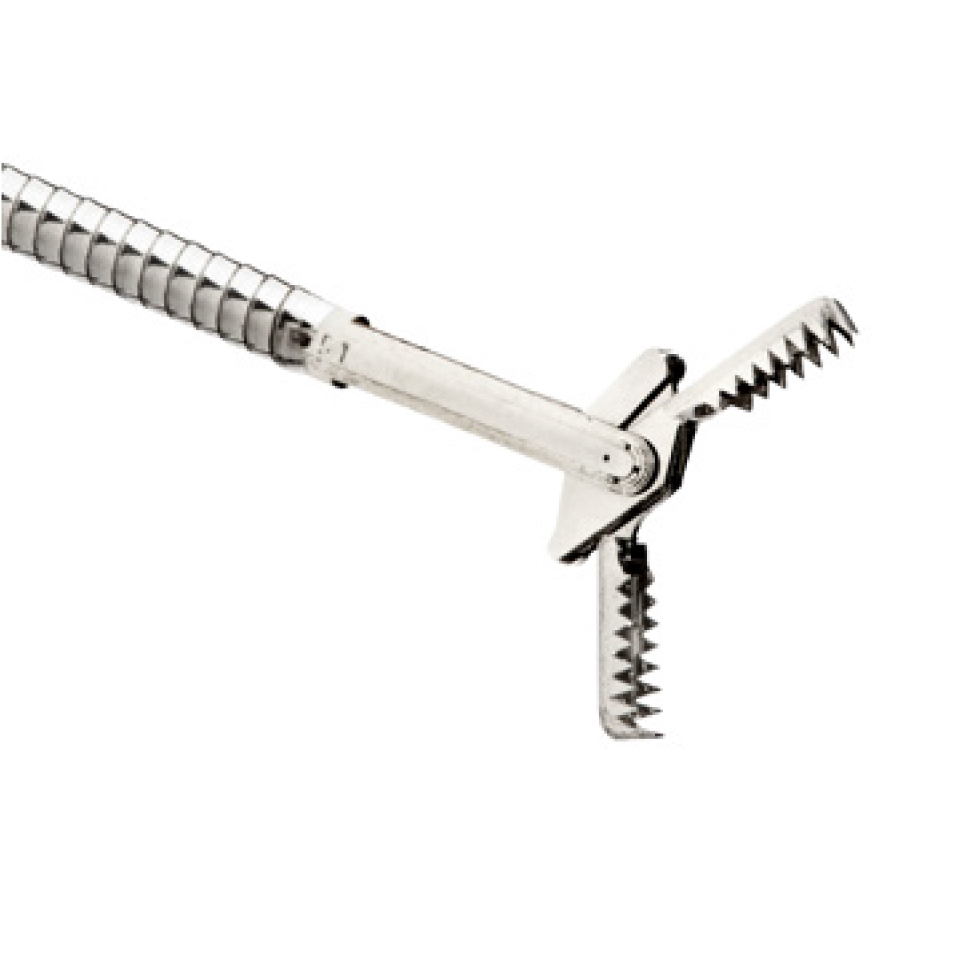
Figure 1 Image of MorayTM microforceps (US Endoscopy, OH, United States).
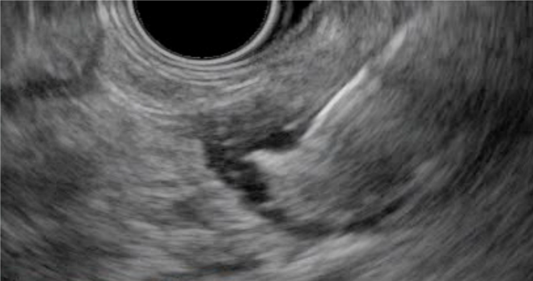
Figure 2 Endosonographic image of the microforceps opened within a pancreatic cystic lesion.
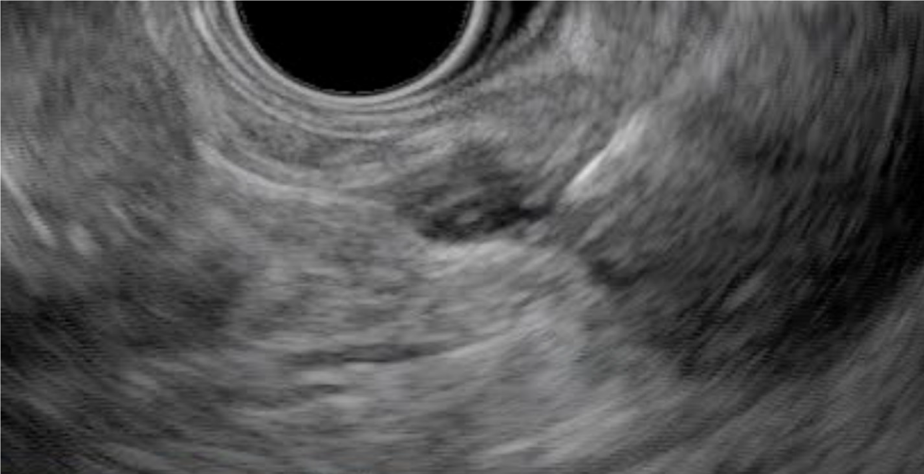
Figure 3 Endosonographic image of the microforceps bite of the wall tenting tissue within a pancreatic cystic lesion.
- Citation: Hashimoto R, Lee JG, Chang KJ, Chehade NEH, Samarasena JB. Endoscopic ultrasound-through-the-needle biopsy in pancreatic cystic lesions: A large single center experience. World J Gastrointest Endosc 2019; 11(11): 531-540
- URL: https://www.wjgnet.com/1948-5190/full/v11/i11/531.htm
- DOI: https://dx.doi.org/10.4253/wjge.v11.i11.531


