Copyright
©The Author(s) 2018.
World J Gastrointest Endosc. Sep 16, 2018; 10(9): 219-224
Published online Sep 16, 2018. doi: 10.4253/wjge.v10.i9.219
Published online Sep 16, 2018. doi: 10.4253/wjge.v10.i9.219
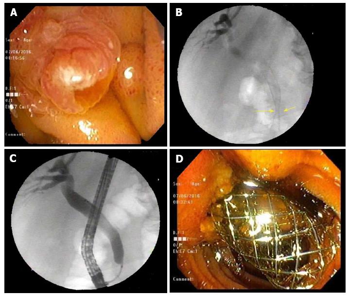
Figure 1 Initial endoscopic retrograde cholangiopancreatography findings.
A: Area of the papilla visualized in the 2nd part of the duodenum; B: CBD malignant stricture visualized in the distal CBD with aid of fluoroscopy during ERCP; C: Cannulating the CBD; D: BMS interested into the CBD visualized protruding from the papilla in the 2nd portion of the duodenum. CBD: Common bile duct; ERCP: Endoscopic retrograde cholangiopancreatography; BMS: Bare metal stent.
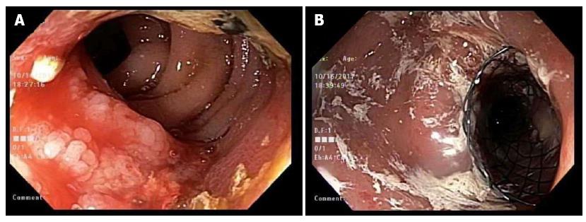
Figure 2 Esophagogastroduodenoscopy findings 15 mo after initial endoscopic retrograde cholangiopancreatography.
A: Duodenal mass visualized encroaching in the lumen of 2nd portion of duodenum; B: SEMS placed over the area of the encroaching mass in the 2nd portion of the duodenum. SEMS: Self expanding metal stent.
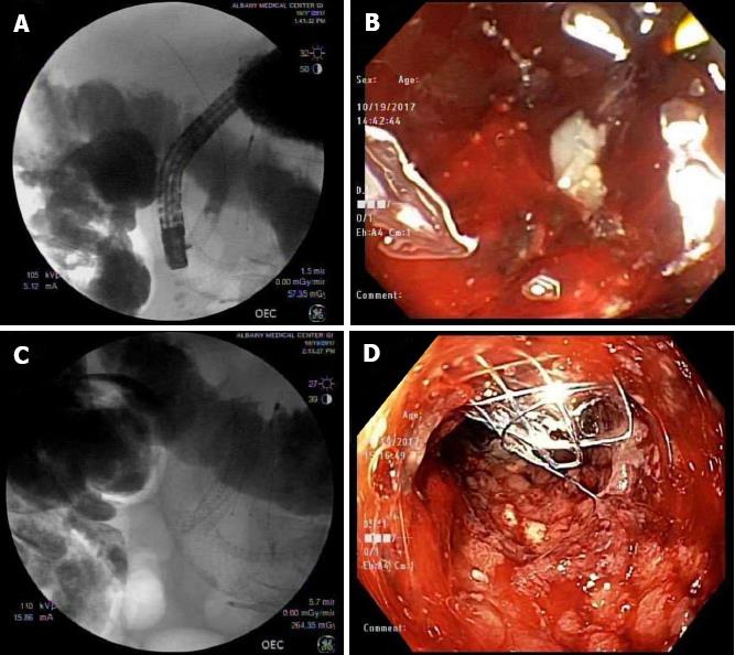
Figure 3 Endoscopic retrograde cholangiopancreatography to place a subsequent 2nd common bile duct stent through an existent duodenal stent.
A: CBD BMS and duodenal prosthesis (SEMS) visualized on the 2nd ERCP before inserting the CMS into the CDB; B: Endoscopic visualization of the papilla site filled with debris and tumor invasion; C: CMS deployed on the existing BMS in the CBD through the SEMS in the duodenum; D: Endoscopic visualization of the CBD stent protruding through the papilla in the 2nd portion of duodenum after the completion of the 2nd ERCP. CBD: Common bile duct; BMS: Bare metal stent; SEMS: Self expanding metal stent; CMS: Covered metal stent; ERCP: Endoscopic retrograde cholangiopancreatography.
- Citation: Virk GS, Parsa NA, Tejada J, Mansoor MS, Hida S. Successful stent-in-stent dilatation of the common bile duct through a duodenal prosthesis, a novel technique for malignant obstruction: A case report and review of literature. World J Gastrointest Endosc 2018; 10(9): 219-224
- URL: https://www.wjgnet.com/1948-5190/full/v10/i9/219.htm
- DOI: https://dx.doi.org/10.4253/wjge.v10.i9.219


