Copyright
©The Author(s) 2018.
World J Gastrointest Endosc. Dec 16, 2018; 10(12): 392-399
Published online Dec 16, 2018. doi: 10.4253/wjge.v10.i12.392
Published online Dec 16, 2018. doi: 10.4253/wjge.v10.i12.392
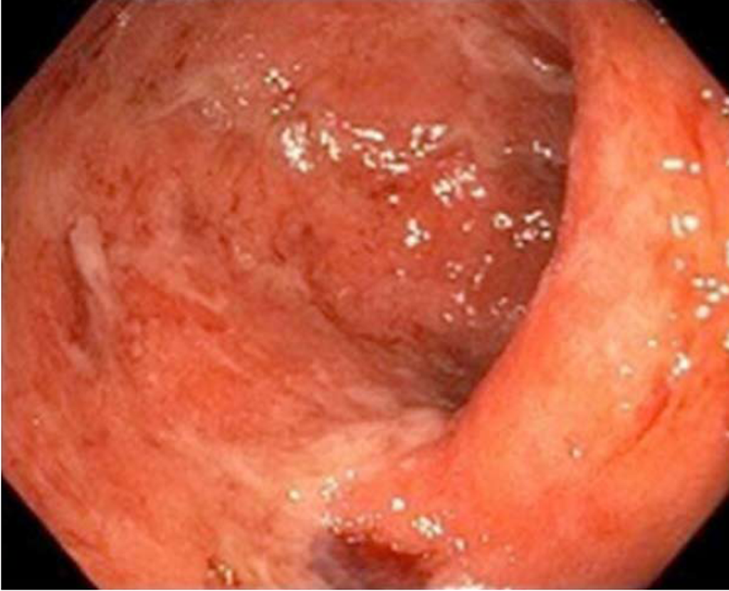
Figure 1 Mucosa at the rectosigmoid junction with mild erythematous spots and no erosions or ulcers.
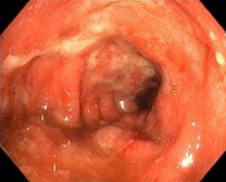
Figure 2 Mucosa at the rectosigmoid junction with erythema and fibrin-covered superficial erosions.
Figure 3 Mucosa in the descending colon with extensive erythema and deep fibrin-covered ulcers.
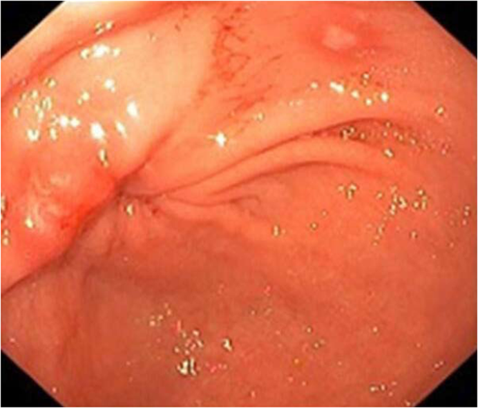
Figure 4 Erosion on the mucosa of the gastric antrum with generalized erythema.
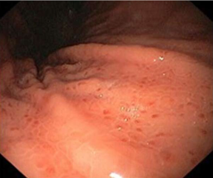
Figure 5 Petechiae on the mucosa of the gastric fold.
- Citation: Iranzo I, Huguet JM, Suárez P, Ferrer-Barceló L, Iranzo V, Sempere J. Endoscopic evaluation of immunotherapy-induced gastrointestinal toxicity. World J Gastrointest Endosc 2018; 10(12): 392-399
- URL: https://www.wjgnet.com/1948-5190/full/v10/i12/392.htm
- DOI: https://dx.doi.org/10.4253/wjge.v10.i12.392


