Copyright
©2009 Baishideng.
World J Gastrointest Endosc. Oct 15, 2009; 1(1): 39-44
Published online Oct 15, 2009. doi: 10.4253/wjge.v1.i1.39
Published online Oct 15, 2009. doi: 10.4253/wjge.v1.i1.39
Figure 1 Images of interventional EUS.
A: EUS FNA with a 22G needle on a solid pancreatic mass; B: CPN: the needle (19G) is inserted immediately adjacent to the lateral aspect of the aorta at the level of the celiac trunk; C: Large pancreatic collection seen from the posterior gastric wall.
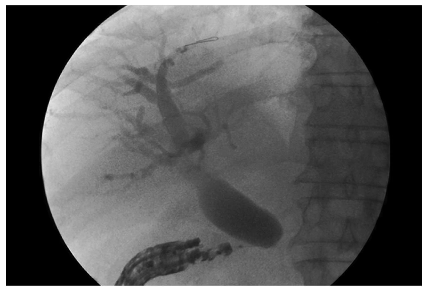
Figure 2 EUS-guided cholangiography through the duodenal wall.
- Citation: Tarantino I, Barresi L. Interventional endoscopic ultrasound: Therapeutic capability and potential. World J Gastrointest Endosc 2009; 1(1): 39-44
- URL: https://www.wjgnet.com/1948-5190/full/v1/i1/39.htm
- DOI: https://dx.doi.org/10.4253/wjge.v1.i1.39


