Copyright
©2009 Baishideng.
World J Gastrointest Endosc. Oct 15, 2009; 1(1): 21-31
Published online Oct 15, 2009. doi: 10.4253/wjge.v1.i1.21
Published online Oct 15, 2009. doi: 10.4253/wjge.v1.i1.21
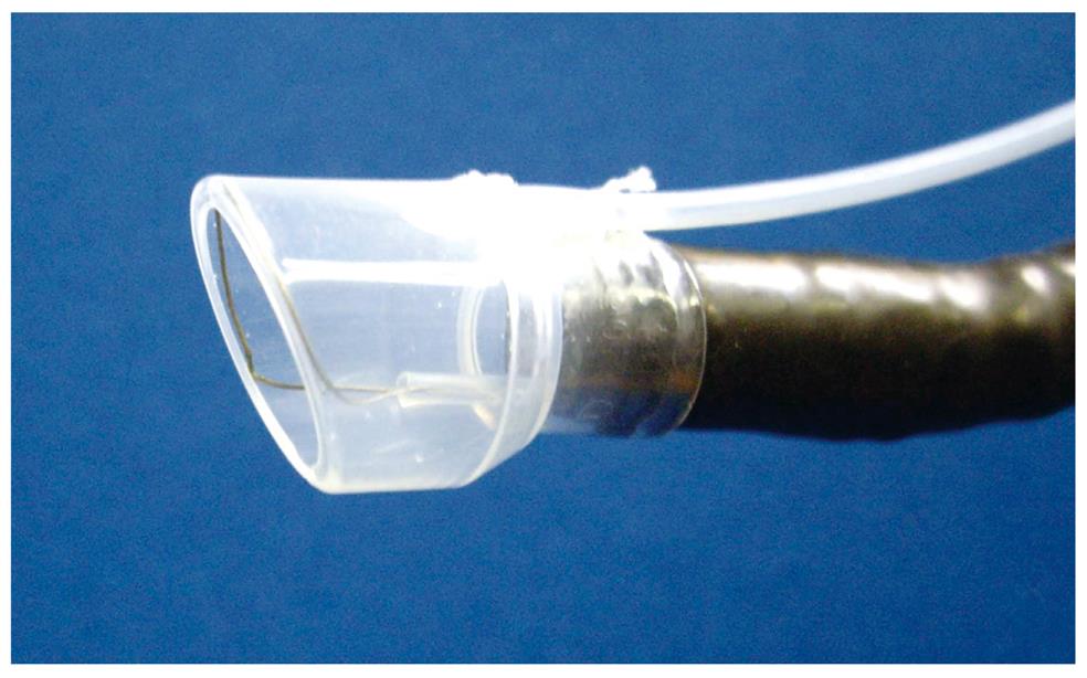
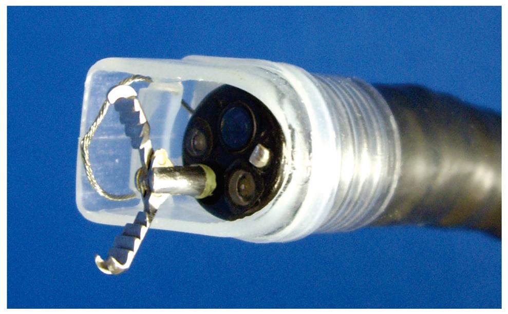
Figure 1 Irrigation prelooped cap.
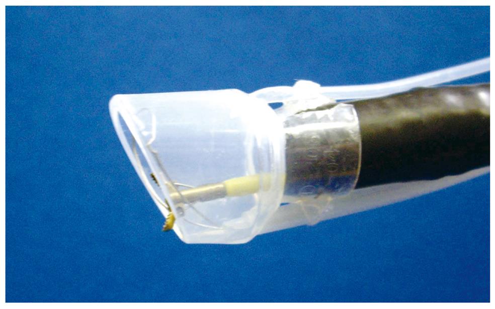
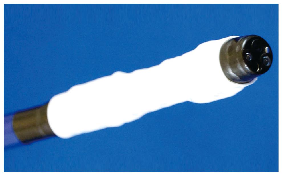
Figure 2 The 2-channel soft prelooped hood.
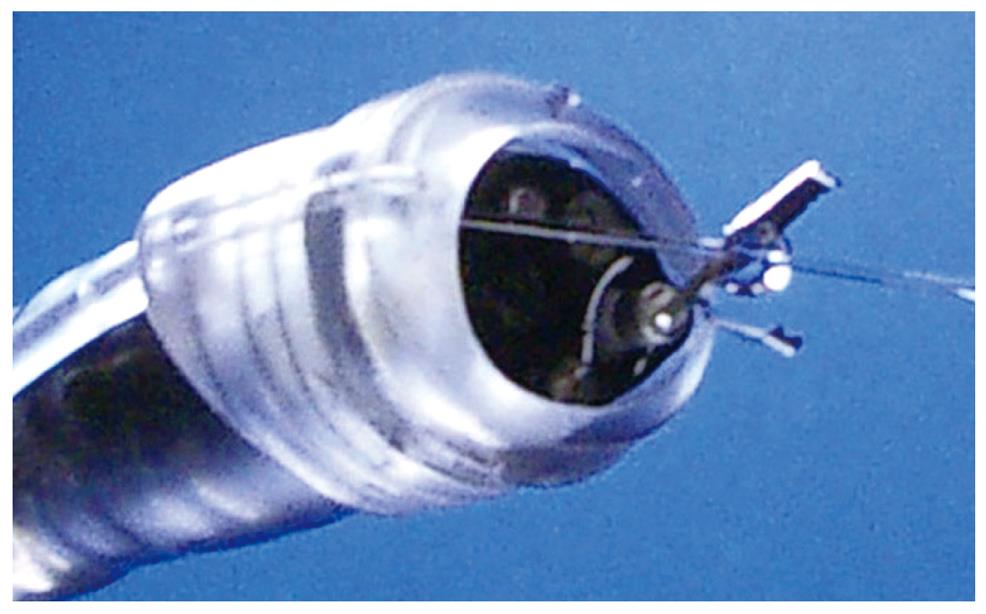
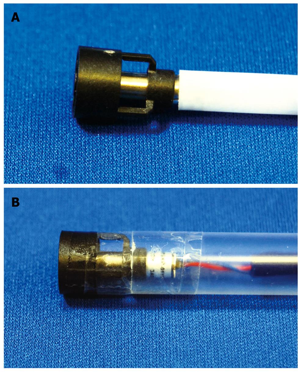
Figure 3 The internally retained snare (IRS) cap.
A: Photograph; B: Schema.
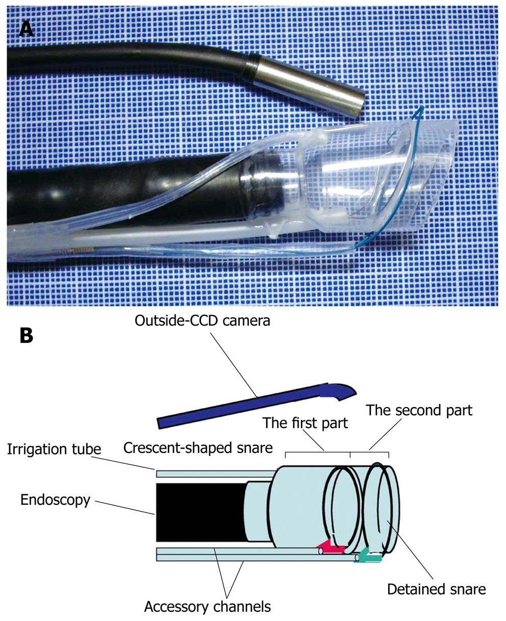
Figure 4 EAMC hood.
A: Placed at the tip of the endoscope and outside-CCD camera; B: Schema of endoscope with EAMC hood. EAMC: endoscopic aspiration mucosectomy and closure.
Figure 5 Vibration hood at the tip of the endoscope.
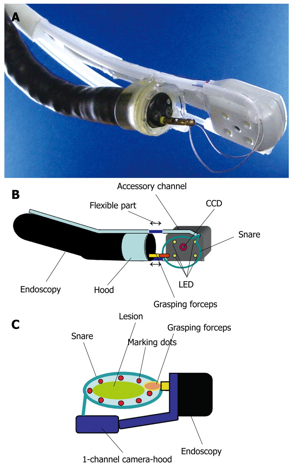
Figure 6 The 1-channel camera-hood.
A: Placed at the tip of the endoscope; B: Schema of the 1-channel camera-hood (B); C: Schematic representation of endoscopic mucosal resection (EMR) using the 1-channel camera-hood.
Figure 7 Irrigation hood knife.
A: Photo; B: Schematic representation of endoscopic submucosal dissection (ESD) using the hood-knife.
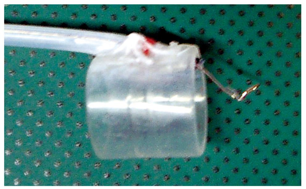
Figure 8 Cap-knife attachment (Type KUME) with a fixed snare placed on the tip of the endoscope through grasping forceps.
Figure 9 The irrigation wiper-knife.
A: Photo; B: Schema of a needle-knife moving like a windshield wiper (both ends red arrow); C: Schematic representation of the ESD using the wiper-knife.
Figure 10 Vibration endoscope.
Figure 11 Irrigation cap (Type KUME) placed on the tip of the endoscope through clipping device.
Figure 12 Coagula-irrigation hood (CI hood).
Figure 13 Endoscopic fan device.
A: Blowing type; B: Ventilation type.
- Citation: Kume K. Endoscopic mucosal resection and endoscopic submucosal dissection for early gastric cancer: Current and original devices. World J Gastrointest Endosc 2009; 1(1): 21-31
- URL: https://www.wjgnet.com/1948-5190/full/v1/i1/21.htm
- DOI: https://dx.doi.org/10.4253/wjge.v1.i1.21


