Published online Aug 27, 2025. doi: 10.4254/wjh.v17.i8.109530
Revised: June 16, 2025
Accepted: July 10, 2025
Published online: August 27, 2025
Processing time: 105 Days and 17.5 Hours
Hepatocellular carcinoma (HCC) is a prevalent and life-threatening cancer with increasing incidence worldwide. High Ki-67 risk stratification is closely associated with higher recurrence rates and worse outcomes following curative therapies in patients with HCC. However, the performance of radiomic and deep transfer learning (DTL) models derived from biparametric magnetic resonance imaging (bpMRI) in predicting Ki-67 risk stratification and recurrence-free survival (RFS) in patients with HCC remains limited.
To develop a nomogram model integrating bpMRI-based radiomic and DTL signatures for predicting Ki-67 risk stratification and RFS in patients with HCC.
This study included 198 patients with histopathologically confirmed HCC who underwent preoperative bpMRI. Ki-67 risk stratification was categorized as high (> 20%) or low (≤ 20%) according to immunohistochemical staining. Radiomic and DTL signatures were extracted from the T2-weighted and arterial-phase images and combined through a random forest algorithm to establish radiomic and DTL models, respectively. Multivariate regression analysis identified clinical risk factors for high Ki-67 risk stratification, and a predictive nomogram model was developed.
A nonsmooth margin and the absence of an enhanced capsule were independent factors for high Ki-67 risk stratification. The area under the curve (AUC) of the clinical model was 0.77, while those of the radiomic and DTL models were 0.81 and 0.87, respectively, for the prediction of high Ki-67 risk stratification, and the nomogram model achieved a better AUC of 0.92. The median RFS times for patients with high and low Ki-67 risk stratification were 33.00 months and 66.73 months, respectively (P < 0.001). Additionally, patients who were predicted to have high Ki-67 risk stratification by the nomogram model had a lower median RFS than those who were predicted to have low Ki-67 risk stratification (33.53 vs 66.74 months, P = 0.007).
Our developed nomogram model demonstrated good performance in predicting Ki-67 risk stratification and predicting survival outcomes in patients with HCC.
Core Tip: In this study, our developed nomogram model can serve as an effective imaging biomarker for predicting Ki-67 risk stratification and predicting survival outcomes in hepatocellular carcinoma patients, with better reliability and clinical utility.
- Citation: Zuo XY, Liu HF. Biparametric magnetic resonance imaging-based radiomic and deep learning models for predicting Ki-67 risk stratification in hepatocellular carcinoma. World J Hepatol 2025; 17(8): 109530
- URL: https://www.wjgnet.com/1948-5182/full/v17/i8/109530.htm
- DOI: https://dx.doi.org/10.4254/wjh.v17.i8.109530
Hepatocellular carcinoma (HCC) is responsible for 75% to 85% of primary liver cancers and is a major contributor to cancer-related illness and death worldwide[1,2]. Despite advances in diagnostic imaging and curative therapies, recurrence remains a substantial threat to the outcomes of patients with HCC[3-5]. The Ki-67 protein serves as a crucial biomarker of cellular proliferation, with high expression levels reflecting tumors with more aggressive biological characteristics[6]. Prior studies have shown that high Ki-67 risk stratification is closely related to increased recurrence rates, greater treatment resistance, and poor survival outcomes in patients with HCC[7-10]. With respect to management strategies, preoperative adjuvant therapy and frequent follow-up are recommended for patients with high Ki-67 risk stratification. Consequently, the assessment of Ki-67 risk stratification would be helpful for individualized therapy planning and survival assessment for patients with HCC.
Radiomics based on magnetic resonance imaging (MRI) has served as a crucial approach for predicting the aggressive features of HCC via the extraction of numerous quantitative signatures[11,12]. Several studies have investigated the efficacy of radiomic models developed on the basis of MRI for the evaluation of Ki-67 risk stratification[13,14], but the corresponding MRI examination is costly and inefficient. Numerous studies have suggested that radiomic models based on T2-weighted imaging (T2WI) combined with arterial phase (AP) imaging have efficiency in predicting aggressive tumor characteristics comparable to those of multisequence MRI[15-17], thus underscoring the importance of biparametric MRI (bpMRI). Additionally, deep transfer learning (DTL) models, implemented through convolutional neural networks, have shown considerable promise in predicting aggressive pathological features[18,19]. However, investigations into both radiomic and DTL models based on bpMRI for predicting Ki-67 risk stratification are lacking, and the majority of previous studies have explored only the ability of MRI-based radiomic models to predict Ki-67 risk stratification[20]. The relationship between radiomic or DTL models in stratifying Ki-67 risk and survival outcomes in HCC patients remains unclear.
Thus, the objective of our study was to develop a nomogram model that integrates bpMRI-based radiomic and DTL signatures for predicting Ki-67 risk stratification and recurrence-free survival (RFS) in HCC patients.
This retrospective study, approved by the Third Affiliated Hospital of Soochow University (Ethical Approval No. 2022-CL027-01) and compliant with the Declaration of Helsinki, involved 218 patients from January 2017 to December 2024. The participants met the following criteria: Histopathologically confirmed HCC post partial hepatectomy, preoperative liver MRI with a 3.0-Tesla scanner, and available Ki-67 index from immunohistochemical analyses. The exclusion criteria were recurrent HCC or other malignancies (n = 8), prior anticancer treatment before MRI (n = 4), severe MRI artifacts or missing sequences (n = 2), and incomplete follow-up data (n = 6). Ultimately, 198 patients were included and randomly assigned to a training group (TG) or a validation group (VG) in a 7:3 ratio.
MRI scans were conducted using a 3.0-Tesla system (Siemens Verio or VIDA, Germany; Phillips Ingenia, Netherlands), and the MRI sequences and imaging parameters are detailed in Supplementary Table 1. For contrast enhancement, a 0.2 mL/kg dose of Gd-DTPA (Magnevist, Beilu, China) was injected at 1 mL/s to capture AP (25-35 second), portal-venous phase (60-70 second), and delayed phase (DP, 180 second) images. The detailed imaging parameters are shown in Supplementary Table 1.
Liver samples were stained with hematoxylin and eosin and then evaluated by an experienced liver pathologist unaware of the patients' preoperative data. Ki-67 risk stratification was measured by the percentage of cells with stained brownish-yellow nuclei: Less than 20%, grade 1 (+); between 20% and 50%, grade 2; and greater than 50%, grade 3 (+++). Furthermore, patients with Ki-67 ≤ 20% were classified into the low-risk subgroup, and those with Ki-67 > 20% were classified into the high-risk subgroup.
Clinical variables, including baseline data, serological results, and MRI semantic features, are shown in Table 1. Notably, to investigate the association between Ki-67 risk stratification and RFS, semantic MRI features were analyzed exclusively for the largest HCC in patients presenting with multiple lesions. Multivariate logistic regression analyses were performed to select risk clinical factors associated with high Ki-67 risk stratification; these factors were then combined via a linear algorithm to develop the clinical model.
| Total (n = 198) | Training (n = 138) | Validation (n = 60) | P value | ||
| Age (Years) | 62.53 ± 10.39 | 62.27 ± 10.38 | 63.13 ± 10.47 | 0.624 | |
| Sex | Male | 164 (82.83) | 117 (84.78) | 47 (78.33) | 0.368 |
| Female | 34 (17.17) | 21 (15.22) | 13 (21.67) | ||
| ALT (U/L) | 35.96 ± 32.93 | 36.51 ± 35.22 | 34.70 ± 27.14 | 0.962 | |
| AST (U/L) | 74.23 ± 44.00 | 71.43 ± 43.04 | 80.65 ± 45.87 | 0.189 | |
| TB (μmol/L) | 16.38 ± 11.32 | 16.35 ± 12.90 | 16.46 ± 6.42 | 0.096 | |
| Cirrhosis | Positive | 55 (27.78) | 39 (28.26) | 16 (26.67) | 0.954 |
| Negative | 143 (72.22) | 99 (71.74) | 44 (73.33) | ||
| AFP | ≤ 20 ng/mL | 106 (53.54) | 77 (55.80) | 29 (48.33) | 0.416 |
| > 20 ng/mL | 92 (46.46) | 61 (44.20) | 31 (51.67) | ||
| HBV/HCV | Positive | 146 (73.74) | 99 (71.74) | 47 (78.33) | 0.428 |
| Negative | 52 (26.26) | 39 (28.26) | 13 (21.67) | ||
| BCLC stage | 0-A | 178 (89.90) | 124 (89.86) | 54 (90.00) | 1.0 |
| BC | 20 (10.10) | 14 (10.14) | 6 (10.00) | ||
| Child-Pugh | A | 183 (92.42) | 125 (90.58) | 58 (96.67) | 0.232 |
| B | 15 (7.58) | 13 (9.42) | 2 (3.33) | ||
| HCC number | 1 | 185 (93.43) | 129 (93.48) | 56 (93.33) | 1.0 |
| 2 | 13 (6.57) | 9 (6.52) | 4 (6.67) | ||
| Irregular shape | Positive | 103 (52.02) | 69 (50.00) | 34 (56.67) | 0.479 |
| Negative | 95 (47.98) | 69 (50.00) | 26 (43.33) | ||
| Nonsmooth margin | Positive | 117 (59.09) | 83 (60.14) | 34 (56.67) | 0.764 |
| Negative | 81 (40.91) | 55 (39.86) | 26 (43.33) | ||
| Enhancing capsule | Positive | 168 (84.85) | 120 (86.96) | 48 (80.00) | 0.299 |
| Negative | 30 (15.15) | 18 (13.04) | 12 (20.00) | ||
| Mosaic architecture | Positive | 115 (58.08) | 84 (60.87) | 31 (51.67) | 0.294 |
| Negative | 83 (41.92) | 54 (39.13) | 29 (48.33) | ||
| Peritumoral enhancement | Positive | 82 (41.41) | 58 (42.03) | 24 (40.00) | 0.913 |
| Negative | 116 (58.59) | 80 (57.97) | 36 (60.00) | ||
| Intratumoral fat content | Positive | 63 (31.82) | 39 (28.26) | 24 (40.00) | 0.143 |
| Negative | 135 (68.18) | 99 (71.74) | 36 (60.00) | ||
| Intratumoral blood products | Positive | 72 (36.36) | 49 (35.51) | 23 (38.33) | 0.827 |
| Negative | 126 (63.64) | 89 (64.49) | 37 (61.67) | ||
| Ki-67 expression | Grade 1 | 119 (60.10) | 78 (56.52) | 41 (68.34) | 0.196 |
| Grade 2 | 53 (26.77) | 42 (30.44) | 11 (18.33) | ||
| Grade 3 | 26 (13.13) | 18 (13.04) | 8 (13.33) | ||
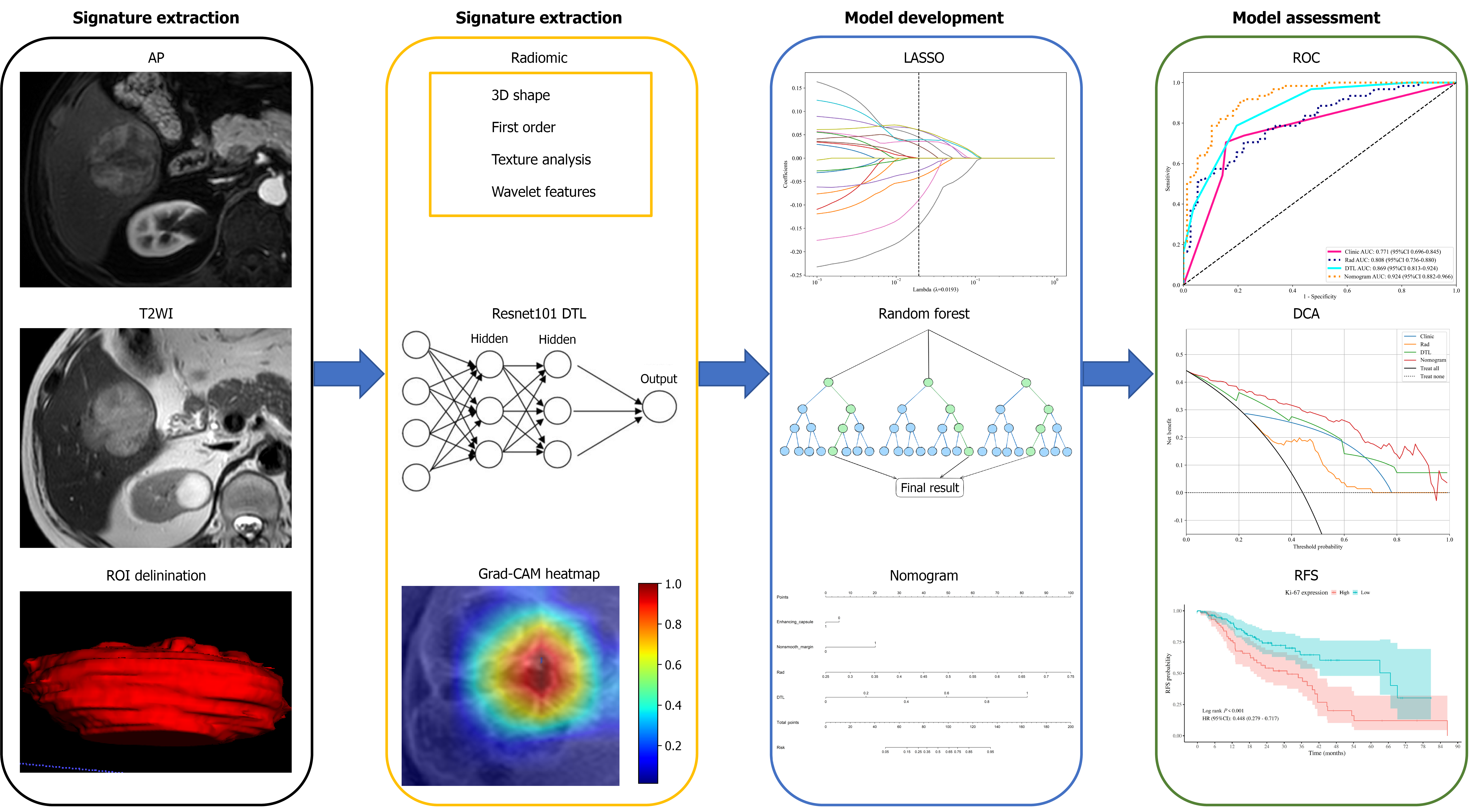
The patients' tumors were manually outlined on T2WI and AP images using ITK-SNAP (version 3.6.0), creating a 3D region of interest (ROI) for the entire HCC. An experienced hepatic radiologist initially delineated the ROI, which was then validated by a senior radiologist. Discrepancies were resolved by consensus between the radiologists, who were unaware of the patients' preoperative clinical and radiological data.
On the basis of the segmented 3D-ROIs, the following sets of radiomic signatures were extracted with the PyRadiomics package (version 3.7.12) implemented in Python: 3D shape (n = 14), first-order (n = 234), texture analysis (n = 222), and wavelet features (n = 728). Consequently, 1198 radiomic signatures were extracted for each T2WI and AP imaging sequence.
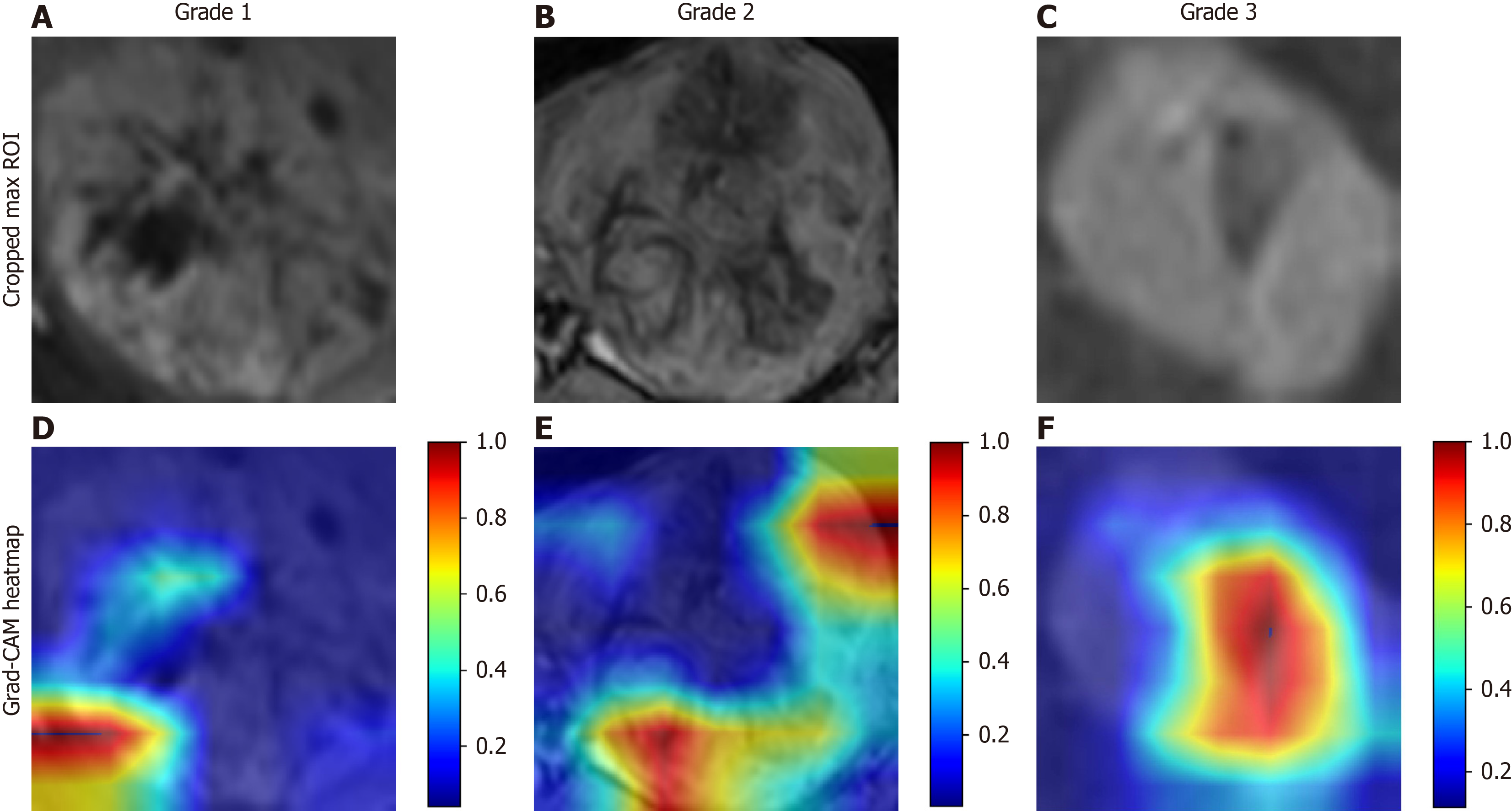
To extract DTL signatures, the ROI with the largest area was cropped and normalized to gray values between 0 and 1 using min-max transformation. The ResNet101 model, a convolutional neural network, was trained on the input data using a cross-entropy loss function, with an adaptive moment estimation optimizer at a 0.1 Learning rate over 500 epochs and a batch size of 50. After training, the model extracted 2048 DTL signatures from the average pooling layer for each HCC lesion in each MRI sequence. Signature visualization was performed using gradient-weighted class activation mapping (Grad-CAM).
A five-step process was used to select robust signatures and develop models: (1) The signature values were normalized using the Z score method; (2) An independent samples t test was used to remove nonsignificant signatures between the high- and low-Ki-67 risk stratification groups; (3) Spearman correlation was applied to identify and retain the better-performing signature from pairs with an absolute correlation > 0.9; (4) LASSO logistic regression with 10-fold cross-validation was used to find robust signatures with nonzero coefficients, which are determined by the optimal penalty parameter (λ); (5) Radiomic and DTL models were developed using a support vector machine (SVM) algorithm, which is based on a linear combination of selected optimal signatures weighted by their coefficients, to predict high Ki-67 risk stratification in HCC.
Following partial hepatectomy, serological examinations and contrast-enhanced MR or computed tomography scans were conducted at regular intervals of 3-6 months. Recurrence was characterized by the emergence of new intrahepatic and/or extrahepatic metastases, as determined by representative radiological findings or pathological results obtained after subsequent surgery. The interval from the initial partial hepatectomy to either the first noted recurrence or the last follow-up was defined as the RFS; patients were censored on March 31, 2025, or at the time of the first recurrence. A comprehensive overview of the process, from MRI acquisition to RFS assessment, is depicted in Figure 1.
Continuous variables are presented as the means ± SDs or medians with interquartile ranges on the basis of the Shapiro-Wilk test results, whereas categorical variables are shown as percentages. Differences between the high and low Ki-67 risk stratification groups were analyzed using the Mann-Whitney U test or χ2 test. Model performance for predicting high Ki-67 risk stratification was assessed using the area under the receiver operating characteristic curve (AUC). Calibration and decision curve analyses (DCAs) were used to evaluate model reliability and clinical utility. RFS curves were created using the Kaplan-Meier method and compared with the log-rank test. All analyses were conducted with R software (version 4.0), with a P value < 0.05 indicating significance.
In this study, a cohort of 198 patients (164 males and 34 females; mean age: 62.53 ± 10.39 years; age range: 29-85 years) was ultimately included and randomly split into either the TG (138 patients) or the VG (60 patients). No significant differences were detected between the TG and VG regarding any baseline clinical factors (P > 0.05), as detailed in Table 1.
According to the immunohistochemical analysis results, 78 (56.52%) patients in the TG and 41 (68.34%) in the VG presented high Ki-67 risk stratification, whereas 60 (43.48%) patients in the TG and 19 (31.66%) in the VG presented low Ki-67 risk stratification (grade 1). Additionally, in the TG and VG, 42 patients (30.44%) and 11 patients (18.33%) were classified as having grade 2 Ki-67 risk stratification, whereas 18 patients (13.04%) and 8 patients (13.33%) were classified as having grade 3 Ki-67 risk stratification.
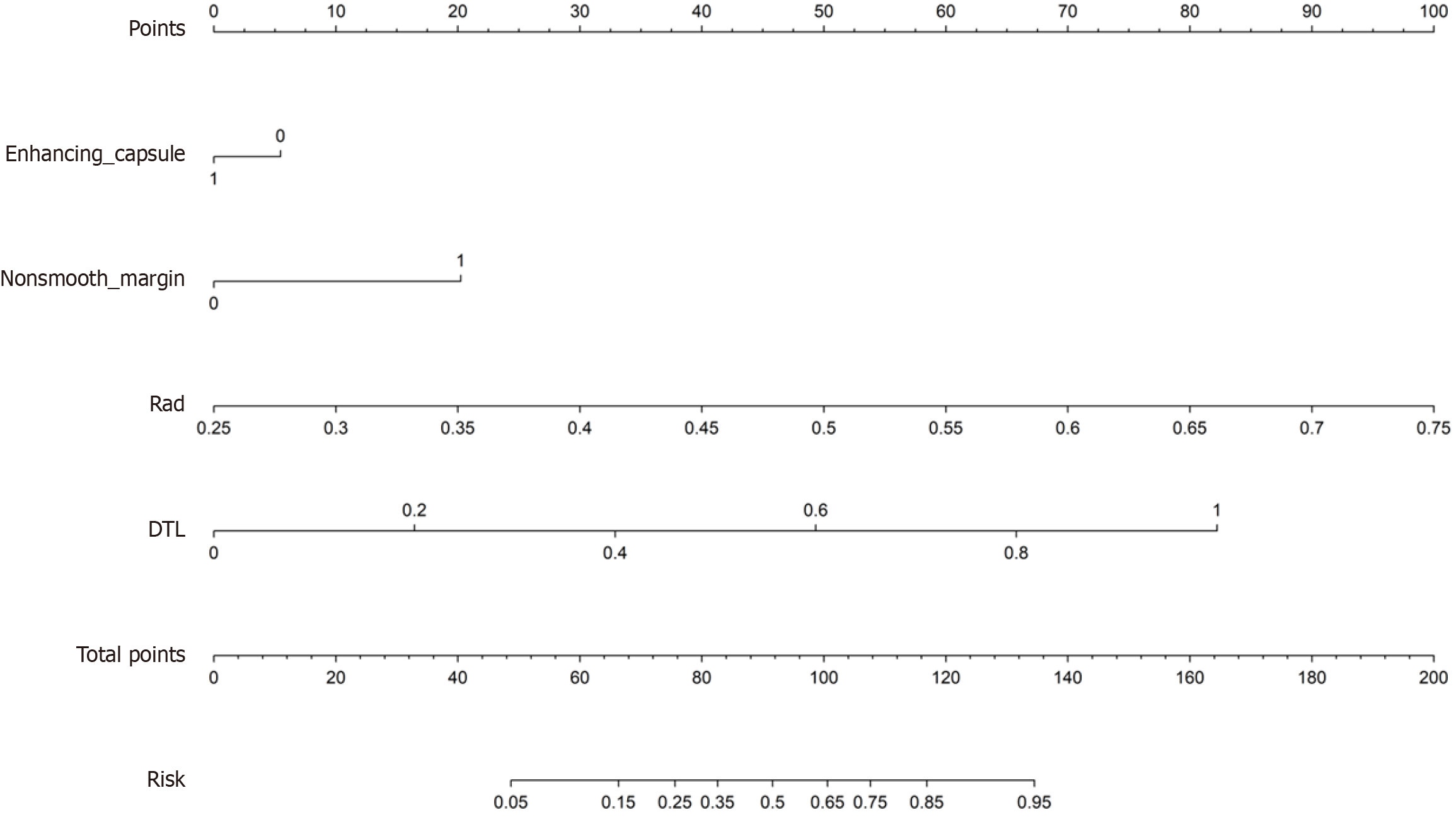
Elevated alpha-fetoprotein (AFP) levels, a nonsmooth margin, and peritumoral enhancement were shown to be significant clinical factors related to high Ki-67 risk stratification (P < 0.05), as summarized in Table 2. Subsequent logistic regression analysis revealed that a nonsmooth margin [odds ratio (OR) = 11.06, 95%CI: 5.60-21.82, P < 0.001] and the absence of an enhanced capsule (OR = 0.27, 95%CI: 0.17-0.42, P < 0.001) were independent risk factors for high Ki-67 risk stratification, as presented in Table 3. The clinical model constructed from these two factors yielded an AUC of 0.77 (95%CI: 0.77-0.85) for TG and 0.72 (95%CI: 0.64-0.88) for VG.
| Training (n = 138) | Validation (n = 60) | ||||||
| High Ki-67 (n = 61) | Low Ki-67 (n = 77) | P value | High Ki-67 (n = 19) | Low Ki-67 (n = 41) | P value | ||
| Age (Years) | 62.42 ± 8.46 | 62.08 ± 12.47 | 0.852 | 61.47 ± 9.64 | 63.90 ± 10.86 | 0.408 | |
| Sex | Male | 53 (86.89) | 64 (83.12) | 0.709 | 16 (84.21) | 31 (75.61) | 0.678 |
| Female | 8 (13.11) | 13 (16.88) | 3 (15.79) | 10 (24.39) | |||
| ALT (U/L) | 34.70 ± 30.76 | 38.80 ± 40.31 | 0.424 | 37.16 ± 21.46 | 33.57 ± 29.59 | 0.137 | |
| AST (U/L) | 66.91 ± 44.02 | 77.15 ± 41.42 | 0.152 | 95.58 ± 37.20 | 73.73 ± 48.23 | 0.066 | |
| TB (μmol/L) | 17.05 ± 15.31 | 15.46 ± 9.05 | 0.85 | 14.56 ± 4.06 | 17.34 ± 7.14 | 0.236 | |
| Tumor size (cm) | 4.47 ± 2.68 | 5.20 ± 3.37 | 0.265 | 6.08 ± 3.56 | 4.88 ± 3.04 | 0.369 | |
| Cirrhosis | Positive | 17 (27.87) | 22 (28.57) | 1.0 | 3 (15.79) | 13 (31.71) | 0.326 |
| Negative | 44 (72.13) | 55 (71.43) | 16 (84.21) | 28 (68.29) | |||
| AFP | ≤ 20 ng/mL | 25 (40.98) | 52 (67.53) | 0.003 | 5 (26.32) | 24 (58.54) | 0.041 |
| > 20 ng/mL | 36 (59.02) | 25 (32.47) | 14 (73.68) | 17 (41.46) | |||
| HBV/HCV | Positive | 44 (72.13) | 55 (71.43) | 1.0 | 16 (84.21) | 31 (75.61) | 0.678 |
| Negative | 17 (27.87) | 22 (28.57) | 3 (15.79) | 10 (24.39) | |||
| BCLC stage | 0-A | 51 (83.61) | 73 (94.81) | 0.06 | 18 (94.74) | 36 (87.80) | 0.711 |
| B-C | 10 (16.39) | 4 (5.19) | 1 (5.26) | 5 (12.20) | |||
| Child-Pugh | A | 55 (90.16) | 70 (90.91) | 1.0 | 18 (94.74) | 40 (97.56) | 1.0 |
| B | 6 (9.84) | 7 (9.09) | 1 (5.26) | 1 (2.44) | |||
| HCC number | 0 | 58 (95.08) | 71 (92.21) | 0.74 | 19 (100.00) | 37 (90.24) | 0.394 |
| 1-2 | 3 (4.92) | 6 (7.79) | 0 (0.00) | 4 (9.76) | |||
| Irregular shape | Positive | 25 (40.98) | 44 (57.14) | 0.087 | 5 (26.32) | 21 (51.22) | 0.126 |
| Negative | 36 (59.02) | 33 (42.86) | 14 (73.68) | 20 (48.78) | |||
| Nonsmooth margin | Positive | 43 (70.49) | 12 (15.58) | < 0.001 | 14 (73.68) | 12 (29.27) | 0.003 |
| Negative | 18 (29.51) | 65 (84.42) | 5 (26.32) | 29 (70.73) | |||
| Enhancing capsule | Positive | 49 (80.33) | 71 (92.21) | 0.071 | 13 (68.42) | 35 (85.37) | 0.238 |
| Negative | 12 (19.67) | 6 (7.79) | 6 (31.58) | 6 (14.63) | |||
| Mosaic architecture | Positive | 29 (47.54) | 25 (32.47) | 0.104 | 9 (47.37) | 20 (48.78) | 1.0 |
| Negative | 32 (52.46) | 52 (67.53) | 10 (52.63) | 21 (51.22) | |||
| Peritumoral enhancement | Positive | 33 (54.10) | 25 (32.47) | 0.017 | 9 (47.37) | 15 (36.59) | 0.61 |
| Negative | 28 (45.90) | 52 (67.53) | 10 (52.63) | 26 (63.41) | |||
| Intratumoral fat content | Positive | 15 (24.59) | 24 (31.17) | 0.508 | 9 (47.37) | 15 (36.59) | 0.61 |
| Negative | 46 (75.41) | 53 (68.83) | 10 (52.63) | 26 (63.41) | |||
| Intratumoral blood products | Positive | 22 (36.07) | 27 (35.06) | 1.0 | 10 (52.63) | 13 (31.71) | 0.206 |
| Negative | 39 (63.93) | 50 (64.94) | 9 (47.37) | 28 (68.29) | |||
| Univariate analysis | Multivariate analysis | |||||
| OR | 95%CI | P value | OR | 95%CI | P value | |
| Age | 0.99 | 0.99-1.00 | 0.170 | |||
| Sex | 0.83 | 0.61-1.12 | 0.310 | |||
| ALT | 0.99 | 0.99-1.00 | 0.609 | |||
| AST | 0.99 | 0.99-1.00 | 0.649 | |||
| TB | 0.99 | 0.97-1.00 | 0.139 | |||
| Cirrhosis | 0.77 | 0.45-1.31 | 0.425 | |||
| AFP | 1.44 | 0.94-2.21 | 0.161 | |||
| HBV/HCV | 0.80 | 0.57-1.12 | 0.270 | |||
| BCLC stage | 2.50 | 0.95-6.61 | 0.121 | |||
| Child-Pugh | 0.86 | 0.34-2.14 | 0.782 | |||
| HCC number | 0.79 | 0.61-1.03 | 0.141 | |||
| Irregular shape | 1.09 | 0.73-1.62 | 0.718 | |||
| Nonsmooth margin | 3.58 | 2.09-6.13 | < 0.001 | 11.06 | 5.60-21.82 | < 0.001 |
| Enhancing capsule | 0.69 | 0.51-0.94 | 0.046 | 0.27 | 0.17-0.42 | < 0.001 |
| Mosaic architecture | 1.16 | 0.74-1.82 | 0.587 | |||
| Intratumoral fat content | 0.63 | 0.36-1.08 | 0.153 | |||
| Intratumoral blood products | 0.82 | 0.51-1.31 | 0.476 | |||
| Peritumoral enhancement | 1.32 | 0.85-2.04 | 0.295 | |||
A total of 2396 radiomic signatures and 4096 DTL signature features were extracted from the biparametric MRI, and their values were subsequently normalized. Following the exclusion of signatures through independent sample t-tests and Spearman correlation analysis, 17 radiomic and 29 DTL signatures were deemed valuable. Among these signatures, 10 optimal radiomic signatures and 25 optimal DTL signatures were identified using the LASSO method, as presented in Supplementary Figures 1-4. The selected signatures were incorporated to develop radiomic and DTL models using the SVM algorithm on the basis of their respective weighted coefficients to predict high Ki-67 risk stratification. The calculated formulas, as well as the corresponding signatures, are illustrated in Supplementary Tables 2 and 3. The cropped maximum ROI and Grad-CAM heatmap of Ki-67 risk stratification ranging from Grade 1 to Grade 3 are shown in Figure 2.
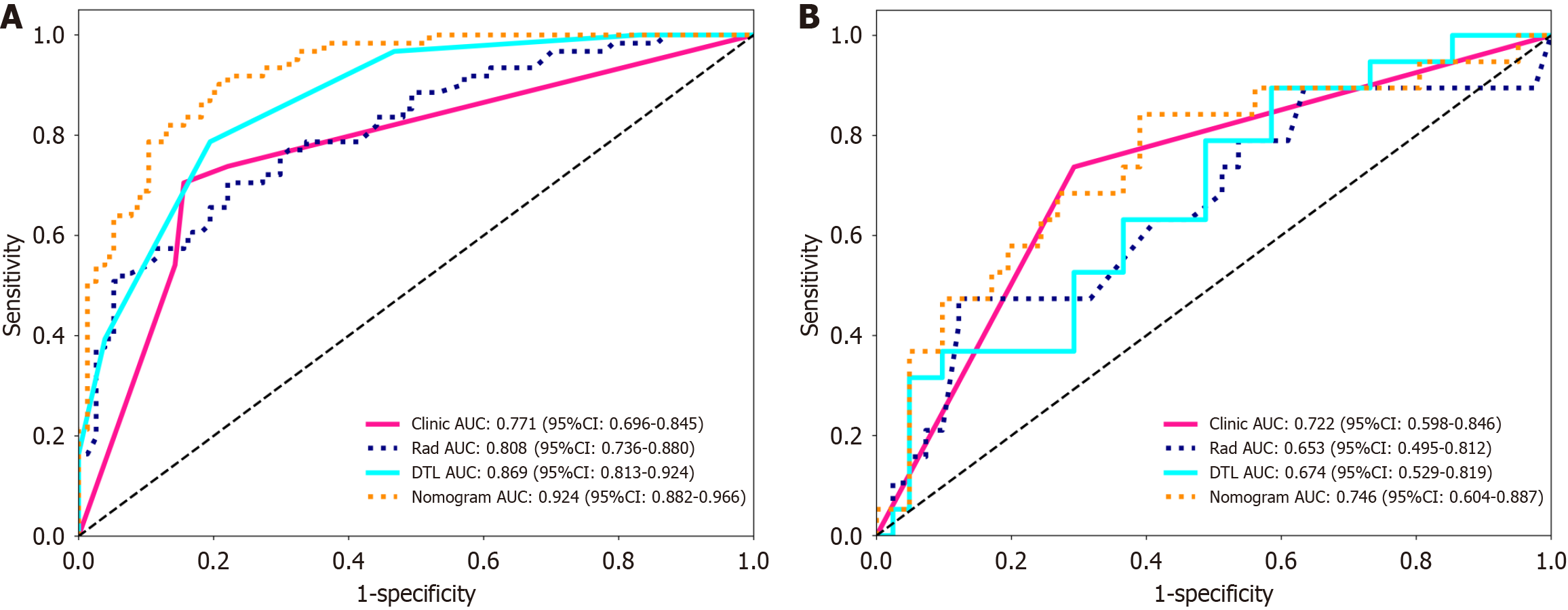
In the prediction of high Ki-67 risk stratification, the radiomic model yielded an AUC value of 0.81 (95%CI: 0.74-0.88) for TG and 0.65 (95%CI: 0.50-0.81) for VG, whereas the DTL model yielded values of 0.87 (95%CI: 0.81-0.92) and 0.67 (95%CI: 0.53-0.82) for TG and VG, respectively, as summarized in Figure 3 and Table 4.
| Model | Number of features | Group | Accuracy | Recall | F1 | AUC (95%CI) | Sensitivity | Specificity |
| Clinical | 2 | Training | 0.78 | 0.705 | 0.741 | 0.77 (0.70-0.84) | 0.71 | 0.84 |
| Validation | 0.72 | 0.737 | 0.622 | 0.72 (0.60-0.85) | 0.74 | 0.71 | ||
| Radiomics | 10 | Training | 0.75 | 0.705 | 0.711 | 0.81 (0.74-0.88) | 0.71 | 0.78 |
| Validation | 0.75 | 0.474 | 0.545 | 0.65 (0.50-0.81) | 0.47 | 0.88 | ||
| DTL | 25 | Training | 0.80 | 0.787 | 0.774 | 0.87 (0.81-0.92) | 0.79 | 0.81 |
| Validation | 0.57 | 0.895 | 0.567 | 0.67 (0.53-0.82) | 0.90 | 0.42 | ||
| Nomogram | 4 | Training | 0.84 | 0.918 | 0.836 | 0.92 (0.88-0.97) | 0.78 | 0.95 |
| Validation | 0.68 | 0.842 | 0.627 | 0.75 (0.60-0.88) | 0.84 | 0.61 |
As illustrated in Figure 4, the nomogram model developed using four significant factors achieved a remarkable AUC value of 0.92 (95%CI: 0.88-0.97) for TG and 0.75 (95%CI: 0.60-0.88) for VG for the evaluation of high Ki-67 risk stratification in HCC patients. Moreover, the nomogram model exhibited significantly superior performance over the other predictive models in terms of TG (P < 0.05). Similarly, the DTL model demonstrated a higher AUC value than did the clinical model; however, no significant differences in AUC values were presented among all the predictive models in the VG (Supplementary Figures 5 and 6).
Calibration curve analysis demonstrated strong reliability between the predicted and histopathologically determined Ki-67 risk stratifications, particularly for the nomogram model. Moreover, DCA plots were generated to graphically illustrate that the nomogram model provided greater net clinical benefits than did the individual predictive models. Notably, all the models exhibited lower reliability and clinical benefits in the VG than in the TG, as depicted in Supplementary Figures 7-10.
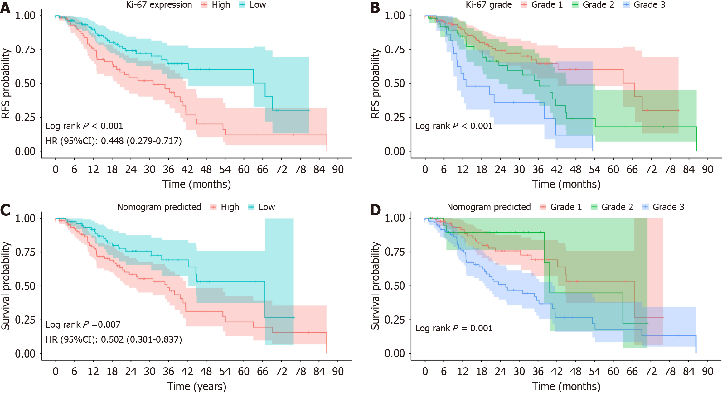
By March 2025, all 198 patients had complete follow-up data, with a median follow-up duration of 17 months (range: 1-86 months). At 1, 3, and 5 years, the RFS rates were 86.87%, 71.21%, and 65.66%, respectively. The median RFS for patients with histopathologically confirmed high (grades 2 and 3) and low (grade 1) Ki-67 risk stratification was 31.00 months (grade 2, 36.33 months; grade 3, 13.03 months) and 66.73 months (P < 0.001), respectively. Additionally, patients predicted by the nomogram to have high Ki-67 risk stratification had a significantly lower median RFS (33.53 months; grade 2, 39.73 months; grade 3, 25.73 months) than those predicted to have low Ki-67 risk stratification (66.74 months; P = 0.007), as shown in Figure 5.
In this study, a novel nomogram model integrating bpMRI-based radiomic and DLT signatures was developed to predict Ki-67 risk stratification and RFS in patients with HCC. The findings demonstrated that both the DLT and radiomic models demonstrated promising value in the evaluation of the risk stratification of Ki-67. Notably, the integration of these models with independent clinical factors into a nomogram model resulted in even greater predictive ability. Furthermore, the findings revealed that patients with either histopathologically diagnosed or nomogram-predicted high Ki-67 risk stratification had a significantly lower median RFS than did those with either actual or predicted low Ki-67 risk stratification following hepatectomy. Additionally, Ki-67 risk stratification emerged as a critical factor influencing RFS in patients with HCC, especially grade 3 Ki-67 risk stratification. Consequently, our study not only develops a noninvasive approach for predicting Ki-67 risk stratification by incorporating clinical and multiscale bpMRI-based radiomic features but also provides more precise information for preoperatively predicting survival outcomes in HCC patients with either histopathologically diagnosed or nomogram-predicted high Ki-67 risk stratification.
Our findings indicate that a nonsmooth margin and the absence of an enhanced capsule are clinical risk factors for high Ki-67 risk stratification, corroborating the conclusions reported by Cai et al[21]. and Liu et al[22]. It is hypothesized that high Ki-67 risk stratification corresponds to active cell proliferation and strong invasive capabilities; thus, related independent clinical factors may serve as indicators of high risk stratification of Ki-67. In contrast to the findings of previous studies[13], elevated AFP levels were not important in distinguishing high vs low Ki-67 risk stratification in this study, possibly because of differences in the study populations. Furthermore, the clinical model presented an AUC value of 0.77, which indicates relatively poor performance in precisely evaluating high Ki-67 risk stratification. Thus, there is an urgent need for more reliable alternatives to predict Ki-67 risk stratification.
Several prior studies have examined the application of MRI-based radiomic models for evaluating Ki-67 risk stratification in HCC, reporting AUCs ranging from 0.77 to 0.80 using a consistent threshold (Ki-67 > 20%)[13,14,20]. Our study revealed a comparable AUC value of 0.81 for the evaluation of high Ki-67 risk stratification on the basis of a bpMRI-based radiomic model. These findings emphasize the efficacy of a radiomic model based on bpMRI (T2WI and AP imaging) as a cost-effective and time-efficient alternative for assessing Ki-67 risk stratification. Furthermore, our radiomic model is more reliable than the clinical model in the prediction of high Ki-67 risk stratification, suggesting that the optimal radiomic signatures extracted from bpMRI offer more detailed insights into the aggressive biological features and intratumoral heterogeneity of HCC[23]. In this study, wavelet heterogeneity on bpMRI was identified as the most influential radiomic signature associated with high Ki-67 risk stratification in HCC, thus suggesting that bpMRI-based wavelet signatures correspond to spatial distributions of proliferating cells in resection specimens, bridging the radiomic model with HCC cellular pathophysiology. DTL has emerged as another promising model for assessing aggressive tumor characteristics and survival outcomes in patients with HCC, yet the performance of an MRI-based DTL model in predicting Ki-67 risk stratification has not been fully investigated. In this research, the DTL model exhibited excellent performance, indicating that the numerous quantitative DTL signatures extracted via a CNN may also serve as a reliable radiomic alternative for the assessment of Ki-67 risk stratification.
Through the promising performance of the bpMRI-based radiomic and DTL models in predicting high Ki-67 risk stratification in patients with HCC, a nomogram model combining multiscale and multimodal features was expected to achieve even greater predictive performance. Thus, we developed a comprehensive nomogram model that ultimately demonstrated superior performance and calibration in predicting high Ki-67 risk stratification, while DCA indicated that the nomogram offered greater clinical benefits than the individual models did, highlighting the added value of integrating radiomic and DTL signatures with clinical factors. Furthermore, the performance of our established nomogram model was greater than those developed in previous studies[13,14], mainly because DTL features were included in establishing the nomogram model. Thus, our findings suggest that the nomogram model is an effective tool for evaluating high Ki-67 risk stratification, facilitating improved clinical decision-making and enabling more precise risk assessment.
Recurrence following hepatectomy is a critical determinant in predicting survival and guiding subsequent therapy in patients with HCC. In our study, the RFS rates at 1, 3, and 5 years were 86.87%, 71.21%, and 65.66%, respectively, which aligns with previous findings[24,25]. Furthermore, HCC patients with high Ki-67 risk stratification, either confirmed via immunohistochemistry or predicted with the nomogram model, had significantly lower median RFS values than did those with low Ki-67 risk stratification. Consequently, high Ki-67 risk stratification was identified as a crucial negative prognostic factor for HCC recurrence after hepatectomy, which is consistent with prior findings[8,9]. These results were attributed to the strong associations between high Ki-67 risk stratification and active cell proliferation and vascular invasion, which likely contribute to a greater incidence of recurrence. Notably, our study further revealed that the median RFS values decreased as the Ki-67 grade increased, emphasizing the importance of risk stratification for Ki-67 risk stratification. Thus, patients with HCC for whom our nomogram model predicted high Ki-67 risk stratification, particularly those with grade 3 risk stratification, should be considered for more active clinical management and frequent monitoring.
This study has several limitations: (1) Although a greater number of patients were included than in prior studies, this retrospective study excluded patients with incomplete follow-up data and lacked external validation, which may pose certain risks of data leakage and overfitting, leading to a loss of model generalizability; (2) To provide a time-efficient alternative and exclude potential data redundancy, radiomic and DTL signatures were extracted solely from bpMRI in our study. Consequently, this approach may overlook promising signatures present in other MRI sequences, including portal venous and delayed phases[26,27], which may provide hallmark features for HCC diagnosis, although bpMRI-based radiomic models have demonstrated approximately equivalent performance in predicting aggressive biological characteristics of HCC compared with multisequence MRI; (3) Although rigorous feature preprocessing was systematically applied to select the most robust radiomic signatures and the SVM algorithm was more suitable for relatively small sample sizes but had more radiomic signatures (n > 1000) than other machine learning algorithms did, the models demonstrated inferior accuracy in the VG than in the TG, mainly because of data overfitting and a relatively small sample size. Thus, future prospective and multicenter studies are essential to improve the predictive model applicability of our research findings; and (4) Due to the limited number of included patients, a 7:3 ratio was used for TG and VG, potentially causing a significant AUC difference between TG and the internal VG among all the predictive models. Although other ratios, such as 8:2 or 6:4, might reduce this difference, the 7:3 ratio is commonly used because it offers enough training data for reliable generalization and is suitable for small samples with many high-dimensional features when constructing radiomic models. Finally, the bpMRI-based radiomic and DTL models improve the predictive power by extracting subvisual features (e.g., fractal dimensions and wavelet-filtered texture patterns) that are undetectable by conventional radiological assessment. However, to enhance predictive model interpretability between the most influential signatures and biological plausibility, implementing SHAP analysis is needed in future research.
Our results highlight that a nomogram model incorporating radiomic and DTL signatures may serve as an effective imaging biomarker for the prediction of Ki-67 risk stratification and survival outcomes in patients with HCC.
| 1. | Bray F, Laversanne M, Sung H, Ferlay J, Siegel RL, Soerjomataram I, Jemal A. Global cancer statistics 2022: GLOBOCAN estimates of incidence and mortality worldwide for 36 cancers in 185 countries. CA Cancer J Clin. 2024;74:229-263. [RCA] [PubMed] [DOI] [Full Text] [Cited by in Crossref: 5690] [Cited by in RCA: 8166] [Article Influence: 8166.0] [Reference Citation Analysis (2)] |
| 2. | Rumgay H, Arnold M, Ferlay J, Lesi O, Cabasag CJ, Vignat J, Laversanne M, McGlynn KA, Soerjomataram I. Global burden of primary liver cancer in 2020 and predictions to 2040. J Hepatol. 2022;77:1598-1606. [RCA] [PubMed] [DOI] [Full Text] [Full Text (PDF)] [Cited by in Crossref: 17] [Cited by in RCA: 1068] [Article Influence: 356.0] [Reference Citation Analysis (0)] |
| 3. | Kim SS, Lee J, Ahn SB, Chon YE, Yoon E, Jeong SW, Jun DW. Clinical Course and Prognosis of Long-Term Survivors of Hepatocellular Carcinoma. Aliment Pharmacol Ther. 2025;61:1333-1342. [RCA] [PubMed] [DOI] [Full Text] [Cited by in Crossref: 1] [Cited by in RCA: 3] [Article Influence: 3.0] [Reference Citation Analysis (0)] |
| 4. | Aziz H, Kwon YIC, Park A, Kwon Y, Aswani Y, Pawlik TM. Comprehensive review of clinical presentation, diagnosis, management, and prognosis of ruptured hepatocellular carcinoma. J Gastrointest Surg. 2024;28:1357-1369. [RCA] [PubMed] [DOI] [Full Text] [Cited by in RCA: 3] [Reference Citation Analysis (0)] |
| 5. | Torimura T, Iwamoto H. Treatment and the prognosis of hepatocellular carcinoma in Asia. Liver Int. 2022;42:2042-2054. [RCA] [PubMed] [DOI] [Full Text] [Cited by in Crossref: 11] [Cited by in RCA: 76] [Article Influence: 25.3] [Reference Citation Analysis (0)] |
| 6. | Karabulut E, Akbulut S, Samdanci ET, Akatli AN, Elsarawy A, Kucukakcali Z, Ogut Z, Tuncer A, Ince V, Yilmaz S. Are Ki-67 and Procalcitonin Expression Levels Useful in Predicting the Biological Behavior of Hepatocellular Carcinoma After Liver Transplantation? J Clin Med. 2024;14:144. [RCA] [PubMed] [DOI] [Full Text] [Full Text (PDF)] [Cited by in RCA: 1] [Reference Citation Analysis (0)] |
| 7. | Xu JX, Qin SL, Wei HW, Chen YY, Peng YC, Qi LN. Prognostic factors and an innovative nomogram model for patients with hepatocellular carcinoma treated with postoperative adjuvant transarterial chemoembolization. Ann Med. 2023;55:2199219. [RCA] [PubMed] [DOI] [Full Text] [Cited by in RCA: 9] [Reference Citation Analysis (0)] |
| 8. | Moriyama M, Kanda T, Midorikawa Y, Matsumura H, Masuzaki R, Nakamura H, Ogawa M, Matsuoka S, Shibata T, Yamazaki M, Kuroda K, Nakayama H, Higaki T, Kanemaru K, Miki T, Sugitani M, Takayama T. The proliferation of atypical hepatocytes and CDT1 expression in noncancerous tissue are associated with the postoperative recurrence of hepatocellular carcinoma. Sci Rep. 2022;12:20508. [RCA] [PubMed] [DOI] [Full Text] [Full Text (PDF)] [Cited by in Crossref: 7] [Cited by in RCA: 9] [Article Influence: 3.0] [Reference Citation Analysis (0)] |
| 9. | Li C, He Z, Hu Y. Is Ki-67 a prognostic factor for post-operation survival in patients with non-alcoholic fatty liver disease-associated hepatocellular carcinoma? Transl Cancer Res. 2022;11:3927-3928. [RCA] [PubMed] [DOI] [Full Text] [Full Text (PDF)] [Cited by in RCA: 1] [Reference Citation Analysis (0)] |
| 10. | Shi Y, Wang X, Zhu Q, Chen G. The Ribosomal Protein L28 Gene Induces Sorafenib Resistance in Hepatocellular Carcinoma. Front Oncol. 2021;11:685694. [RCA] [PubMed] [DOI] [Full Text] [Full Text (PDF)] [Cited by in RCA: 9] [Reference Citation Analysis (0)] |
| 11. | Liu HF, Zhang YZ, Wang Q, Zhu ZH, Xing W. A nomogram model integrating LI-RADS features and radiomics based on contrast-enhanced magnetic resonance imaging for predicting microvascular invasion in hepatocellular carcinoma falling the Milan criteria. Transl Oncol. 2023;27:101597. [RCA] [PubMed] [DOI] [Full Text] [Full Text (PDF)] [Cited by in Crossref: 9] [Cited by in RCA: 12] [Article Influence: 6.0] [Reference Citation Analysis (0)] |
| 12. | Liu HF, Lu Y, Wang Q, Lu YJ, Xing W. Machine Learning-Based CEMRI Radiomics Integrating LI-RADS Features Achieves Optimal Evaluation of Hepatocellular Carcinoma Differentiation. J Hepatocell Carcinoma. 2023;10:2103-2115. [RCA] [PubMed] [DOI] [Full Text] [Full Text (PDF)] [Cited by in Crossref: 11] [Cited by in RCA: 12] [Article Influence: 6.0] [Reference Citation Analysis (0)] |
| 13. | Yan Y, Lin XS, Ming WZ, Chuan ZQ, Hui G, Juan SY, Shuang W, Yang Fan LV, Dong Z. Radiomic Analysis Based on Gd-EOB-DTPA Enhanced MRI for the Preoperative Prediction of Ki-67 Expression in Hepatocellular Carcinoma. Acad Radiol. 2024;31:859-869. [RCA] [PubMed] [DOI] [Full Text] [Cited by in Crossref: 1] [Cited by in RCA: 6] [Article Influence: 6.0] [Reference Citation Analysis (0)] |
| 14. | Fan Y, Yu Y, Wang X, Hu M, Hu C. Radiomic analysis of Gd-EOB-DTPA-enhanced MRI predicts Ki-67 expression in hepatocellular carcinoma. BMC Med Imaging. 2021;21:100. [RCA] [PubMed] [DOI] [Full Text] [Full Text (PDF)] [Cited by in Crossref: 6] [Cited by in RCA: 33] [Article Influence: 8.3] [Reference Citation Analysis (0)] |
| 15. | Yang C, Zhang ZM, Zhao ZP, Wang ZQ, Zheng J, Xiao HJ, Xu H, Liu H, Yang L. Radiomic analysis based on magnetic resonance imaging for the prediction of VEGF expression in hepatocellular carcinoma patients. Abdom Radiol (NY). 2024;49:3824-3833. [RCA] [PubMed] [DOI] [Full Text] [Full Text (PDF)] [Cited by in Crossref: 4] [Cited by in RCA: 6] [Article Influence: 6.0] [Reference Citation Analysis (0)] |
| 16. | Li SQ, Yang CX, Wu CM, Cui JJ, Wang JN, Yin XP. Prediction of glypican-3 expression in hepatocellular carcinoma using multisequence magnetic resonance imaging-based histology nomograms. Quant Imaging Med Surg. 2024;14:4436-4449. [RCA] [PubMed] [DOI] [Full Text] [Full Text (PDF)] [Cited by in RCA: 1] [Reference Citation Analysis (0)] |
| 17. | Gao L, Xiong M, Chen X, Han Z, Yan C, Ye R, Zhou L, Li Y. Multi-Region Radiomic Analysis Based on Multi-Sequence MRI Can Preoperatively Predict Microvascular Invasion in Hepatocellular Carcinoma. Front Oncol. 2022;12:818681. [RCA] [PubMed] [DOI] [Full Text] [Full Text (PDF)] [Cited by in RCA: 15] [Reference Citation Analysis (0)] |
| 18. | Xia T, Zhao B, Li B, Lei Y, Song Y, Wang Y, Tang T, Ju S. MRI-Based Radiomics and Deep Learning in Biological Characteristics and Prognosis of Hepatocellular Carcinoma: Opportunities and Challenges. J Magn Reson Imaging. 2024;59:767-783. [RCA] [PubMed] [DOI] [Full Text] [Cited by in Crossref: 8] [Cited by in RCA: 35] [Article Influence: 35.0] [Reference Citation Analysis (0)] |
| 19. | Liu HF, Wang M, Lu YJ, Wang Q, Lu Y, Xing F, Xing W. CEMRI-Based Quantification of Intratumoral Heterogeneity for Predicting Aggressive Characteristics of Hepatocellular Carcinoma Using Habitat Analysis: Comparison and Combination of Deep Learning. Acad Radiol. 2024;31:2346-2355. [RCA] [PubMed] [DOI] [Full Text] [Cited by in RCA: 14] [Reference Citation Analysis (0)] |
| 20. | Zhou L, Chen Y, Li Y, Wu C, Xue C, Wang X. Diagnostic value of radiomics in predicting Ki-67 and cytokeratin 19 expression in hepatocellular carcinoma: a systematic review and meta-analysis. Front Oncol. 2023;13:1323534. [RCA] [PubMed] [DOI] [Full Text] [Cited by in RCA: 5] [Reference Citation Analysis (0)] |
| 21. | Cai C, Wang L, Tao L, Zhu H, Ren Y, Li D, Li D. Imaging-Based Prediction of Ki-67 Expression in Hepatocellular Carcinoma: A Retrospective Study. Cancer Med. 2025;14:e70562. [RCA] [PubMed] [DOI] [Full Text] [Cited by in RCA: 1] [Reference Citation Analysis (0)] |
| 22. | Liu Z, Yang S, Chen X, Luo C, Feng J, Chen H, Ouyang F, Zhang R, Li X, Liu W, Guo B, Hu Q. Nomogram development and validation to predict Ki-67 expression of hepatocellular carcinoma derived from Gd-EOB-DTPA-enhanced MRI combined with T1 mapping. Front Oncol. 2022;12:954445. [RCA] [PubMed] [DOI] [Full Text] [Full Text (PDF)] [Cited by in RCA: 10] [Reference Citation Analysis (0)] |
| 23. | Nie P, Zhang J, Miao W, Duan S, Wang T, Zhang J, Gu J, Wang N, Zhang R, Wang X, Yang G, Rao W, Wang Z. Incremental value of radiomics-based heterogeneity to the existing risk criteria in predicting recurrence of hepatocellular carcinoma after liver transplantation. Eur Radiol. 2023;33:6608-6618. [RCA] [PubMed] [DOI] [Full Text] [Cited by in Crossref: 2] [Cited by in RCA: 7] [Article Influence: 3.5] [Reference Citation Analysis (0)] |
| 24. | Drefs M, Schoenberg MB, Börner N, Koliogiannis D, Koch DT, Schirren MJ, Andrassy J, Bazhin AV, Werner J, Guba MO. Changes of long-term survival of resection and liver transplantation in hepatocellular carcinoma throughout the years: A meta-analysis. Eur J Surg Oncol. 2024;50:107952. [RCA] [PubMed] [DOI] [Full Text] [Cited by in RCA: 10] [Reference Citation Analysis (0)] |
| 25. | Li H, Meng L, Yu S, Zheng H, Yu L, Wang H, Ren H, Li H, Zhang X, Wang Z, Yu P, Hu X, Yang M, Yan J, Shao Y, Cao L, Ding X, Hong Z, Zhu Z. Efficacy and safety of robotic versus laparoscopic liver resection for hepatocellular carcinoma: a propensity score-matched retrospective cohort study. Hepatol Int. 2024;18:1271-1285. [RCA] [PubMed] [DOI] [Full Text] [Cited by in Crossref: 1] [Cited by in RCA: 8] [Article Influence: 8.0] [Reference Citation Analysis (0)] |
| 26. | Matsuda K, Ueno A, Tsuzaki J, Kurebayashi Y, Masugi Y, Yamazaki K, Tamura M, Abe Y, Hasegawa Y, Kitago M, Jinzaki M, Sakamoto M. Vessels encapsulating tumor clusters contribute to the intratumor heterogeneity of HCC on Gd-EOB-DTPA-enhanced MRI. Hepatol Commun. 2025;9:e0593. [RCA] [PubMed] [DOI] [Full Text] [Cited by in RCA: 4] [Reference Citation Analysis (0)] |
| 27. | Zhang Y, Chen J, Yang C, Dai Y, Zeng M. Preoperative prediction of microvascular invasion in hepatocellular carcinoma using diffusion-weighted imaging-based habitat imaging. Eur Radiol. 2024;34:3215-3225. [RCA] [PubMed] [DOI] [Full Text] [Cited by in Crossref: 1] [Cited by in RCA: 10] [Article Influence: 10.0] [Reference Citation Analysis (0)] |


