Published online Jan 27, 2022. doi: 10.4254/wjh.v14.i1.98
Peer-review started: May 22, 2021
First decision: July 6, 2021
Revised: July 17, 2021
Accepted: November 30, 2022
Article in press: November 30, 2021
Published online: January 27, 2022
Processing time: 243 Days and 21.5 Hours
Recent evidence points towards the role of genotype to understand the phenotype, predict the natural course and long term outcome of patients with progressive familial intrahepatic cholestasis (PFIC). Expanded role of the heterozygous transporter defects presenting late needs to be suspected and identified. Treatment of pruritus, nutritional rehabilitation, prevention of fibrosis pro
Core Tip: The spectrum of clinical manifestations in progressive familial intrahepatic cholestasis varies from mild to severe leading to end stage liver disease necessitating liver transplantation. Medical therapy forms the mainstay of treatment of pruritus with surgical biliary diversion reserved for refractory cases. Apical sodium bile salt co-transporter inhibitors are among the most promising newer drugs. This article describes the recent advances in understanding the clinical course and emerging therapies.
- Citation: Alam S, Lal BB. Recent updates on progressive familial intrahepatic cholestasis types 1, 2 and 3: Outcome and therapeutic strategies. World J Hepatol 2022; 14(1): 98-118
- URL: https://www.wjgnet.com/1948-5182/full/v14/i1/98.htm
- DOI: https://dx.doi.org/10.4254/wjh.v14.i1.98
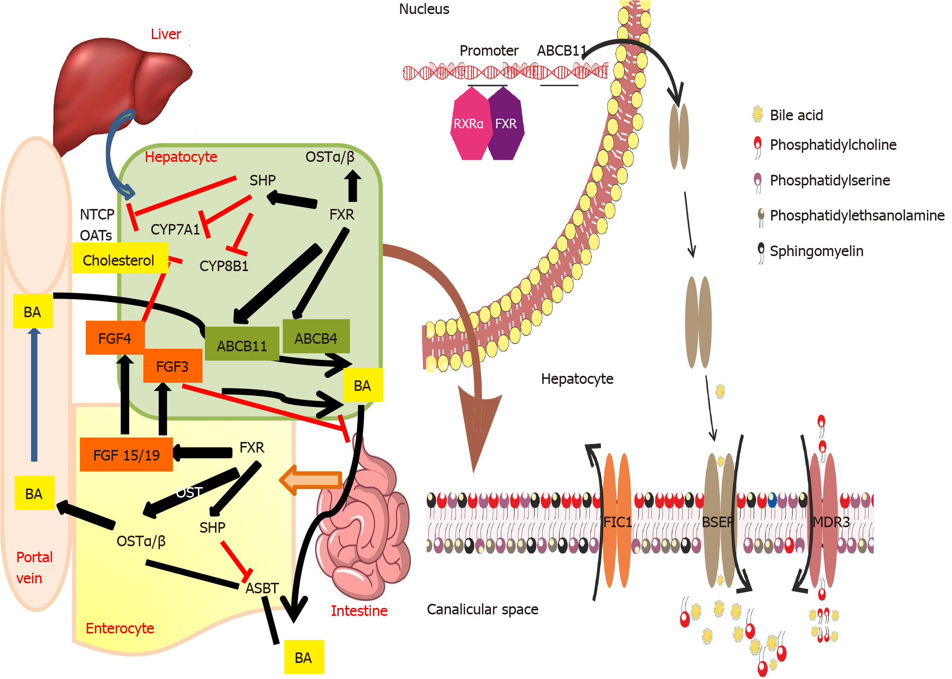
Progressive familial intrahepatic cholestasis (PFIC) is estimated to affect 1 in 50000-100000 births[1]. With improved awareness about the various presentations of these bile transport disorders, we now know that the clinical presentation can vary from early-onset severe liver disease to episodic late-onset occurrence triggered by an external stimulus. Figure 1 describes the various transporters which are involved in different types of PFIC. This article will focus on the PFIC types 1, 2 and 3. Table 1 depicts the phenotype and genotype of these three types of PFIC.
| PFIC1 | PFIC2 | PFIC 3 | |
| Locus/gene/protein | 18q21-22/ATP8B1/FIC1 | 2q24/ABCB11/BSEP | 7q21/ABCB4/MDR3 |
| Known mutations (n)1 | 50 | 200 | 300 |
| Clinical profile | |||
| Onset | Early onset | Early onset | Second decade |
| Age of presentation pruritus | 60% by 3 mo | 72% by 3 mo | 2-3 yr |
| Jaundice | Severe | Severe | Mild to none |
| Cirrhosis | Severe; By end of first decade | Severe; Majority within first 2 yr of life | Mild to moderate; By end of first decade |
| Growth failure | Present 90% | Present 59% | |
| Others | Diarrhea 61%; Pneumonia 13%; Pancreatitis 12%; Deafness 31% | Gall stones in 32% | Delayed puberty |
| Progression | Moderate rate of progression | Rapidly progressive | Highly variable rate of progression |
| Associations with other cholestatic presentations | BRIC; ICP | BRIC, DIC; ICP, HCC | DIC, LPAC; ICP |
| Laboratory profile | |||
| TBA | High | Very high | High |
| GGT | Low to normal | Low to normal | High |
| AST/ALT | Mild elevation | Moderate elevation | Mild elevation |
| AFP | Normal | High | Normal |
| Histopathology | As disease progresses, periportal & pericentrilobular fibrosis develops; Leads to bridging fibrosis and micronodular cirrhosis | Canalicular cholestasis, lobular/portal fibrosis and inflammation with giant cells; Severe hepatocellular necrosis | Portal inflammation, portal fibrosis, cholestasis, ductular proliferation |
| Immunohistochemistry | Canalicular BSEP is normal or faint and MDR3 is normal bland intralobular cholestasis | BSEP expression decreased to absent in the canalicular membrane | MDR3 decreased to absent in the canalicular membrane |
Prevalence of familial intrahepatic cholestasis 1 (FIC1) deficiency is 10.4%-37.5% amongst the cholestatic disorders[2]. Earlier known as Byler’s disease, seen predominantly in children of Amish descent, this disease is now known to occur in all racial and ethnic groups.
FIC1 is encoded by the ATPase phospholipid transporting-8B1 gene (ATP8B1), located on chromosome 18 and responsible for maintaining the asymmetric distribution of phospholipids across the lipid bilayer by the flippase movement of phospholipids from the outer lipid leaflet of the canalicular membrane to the inner lipid leaflet. Earlier it was believed that FIC1 was responsible for the flippase movement of phosphatidyl-serine, but it is now proven that the preferred substrate is phosphatidyl-choline[3]. Phosphatidyl-choline helps in protecting the canalicular membrane from hydrophobic bile acids (BA). Hence, in the absence of FIC1, proteins in the canalicular membrane such as the bile salt export pump (BSEP) can have impaired function. ATP-binding cassette subfamily B member 4 (ABCB4) and ATP8B1 maintain lipid asymmetry which is important for the integrity of the canalicular membrane. When ABCB4 flops the phosphatidyl-choline, there is destabilization of the canalicular membrane which is counteracted by the flippase activity of FIC1. Hence by main
FIC1 is expressed in a variety of tissues, including the canalicular membrane of hepatocytes, apical membrane of cholangiocytes, brush border of enterocytes and cochlear hair cells[9]. Hearing loss is attributed to defects in the composition of membranes of inner ear cilia. Knockdown ATP8B1 in Caco-2 cell model of intestinal epithelium leads to an unorganized apical actin cytoskeleton and post-transcriptional defect in apical protein expression. This prevents the movement of the ATP8B1 into the apical membrane further causing inhibition of ASBT through the intestinal FXR, eventually resulting in bile malabsorption and diarrhea[8]. FIC1 is also expressed in the pneumocytes where ATP8B1 inhibits cardiolipin. Hence deficiency of ATP8B1 increases the cardiolipin which can disrupt surfactant function within pulmonary alveoli and increase the risk for pneumonia[10]. ATP8B1 deficiency is also associated with down-regulation of cystic fibrosis transmembrane conductance regulator (CFTR) and increased sweat chloride test in 15% of patients.
Based on the patients’ clinical, laboratory profile, and liver histology, the presentation of ATP8B1 deficiency ranges from a severe (earlier known as PFIC1) to a milder phenotype [earlier known as benign recurrent intrahepatic cholestasis (BRIC)][11].
Severe ATP8B1 deficiency presents in children with refractory cholestasis (typically beginning in early infancy) manifesting as jaundice, severe failure to thrive (disproportionate to the degree of cholestasis), delayed puberty and fat-soluble vitamin defi
Mild ATP8B1 deficiency: Episodic cholestasis beginning later in childhood, ado
In a recent study with 18 potential disease-causing mutations in ATP8B1, 14 patients (4 homozygous, 6 compound heterozygous, 4 heterozygous) were below 18 years of age and had a median age of 0.75 years[16]. Overall, 28 different genetic variants were identified including two common single nucleotide polymorphisms (SNPs) (p.R952Q and c.3531+8G>T)[16]. These variants have been described earlier in European patients with intrahepatic cholestasis of pregnancy (ICP)[17] and pancreatitis[13]. Pathogenic variants with underlying severe ATP8B1 deficiency are likely to be fully penetrant but some of the milder variants may not become symptomatic even in adulthood. The p.Ile661Thr pathogenic variant, which is frequently detected in people with a mild disease of European descent, appears occasionally to be non-penetrant. However, it is also found in compound heterozygous form in persons with severe disease. A mul
The majority of the severe variants of ATP8B1 deficiency will have disease onset within infancy. Some may experience episodes of severe cholestasis followed by disease-free intervals with eventual persistence of cholestasis. Malnutrition and fat-soluble vitamin deficiencies can cause significant morbidity, if left untreated. Features of chronic liver disease and portal hypertension may develop leading to cirrhosis by the end of the first decade of life but variations have been noted within families[15]. Without surgical intervention, end-stage hepatic failure and death usually occur in the second decade of life. In some of those with mild disease, clinical monitoring may reveal a shift in the clinical spectrum from mild intermittent disease to more persistent cholestasis and fibrosis on the follow-up biopsies[2]. The Natural course and Prognosis of PFIC and Effect of biliary Diversion (NAPPED) consortium data revealed that 8 of 130 patients (6%) died before liver transplantation (LT), of which 3 had undergone surgical biliary diversion (SBD)[15]. Survival analysis showed that at 18 years of age, 44% of patients were alive with their native liver. Three-fourths of those alive with their native liver had undergone SBD by 18 years and a significantly lower percentage of patients with a FIC1-C genotype underwent SBD (FIC1-A 65%, FIC1-B 57%, FIC1-C 45%; P = 0.03). Native liver survival (NLS) was comparable between the three genotype sub-groups at 10 years of age (FIC1-A 67%, FIC1-B 41%, FIC1-C 59%; P = 0.15) with or without SBD. However, the proportions of total patients alive with the native liver at the age of 10 years were lower in patients without SBD than in patients who had undergone an SBD during follow-up.
BSEP is a liver-specific transporter involved in actively transporting monovalent bile salts out of hepatocytes into biliary canaliculi against a concentration gradient. BSEP is encoded by the ABCB11 gene located on chromosome 2q31. Homozygous or com
Chenodeoxycholic acid (CDCA) and the Retinoid X Receptors (FXR) α-heterodimer together activate the ABCB11[18]. Increasing bile acid concentration in the hepatocytes stimulates greater synthesis of BSEP to maintain equilibrium. There are over 200 mutations of ABCB11 causing PFIC2[18]. Common mutants that cause retention of the protein in the endoplasmic reticulum are G238V, D482G, G982R, R1153C, R1286Q, and ΔGly. The mutant protein that is retained and translocated out of the endoplasmic reticulum is then broken down by proteasomes[19]. Intrahepatic accumulation of bile salts and inflammation promote carcinogenesis through genomic modifications[19]. Metabolomic studies have shown that ABCB11 knock-out mice have impaired mitochondrial fatty acid β-oxidation with the resulting increase in reactive oxygen species that might exacerbate the liver injury[19].
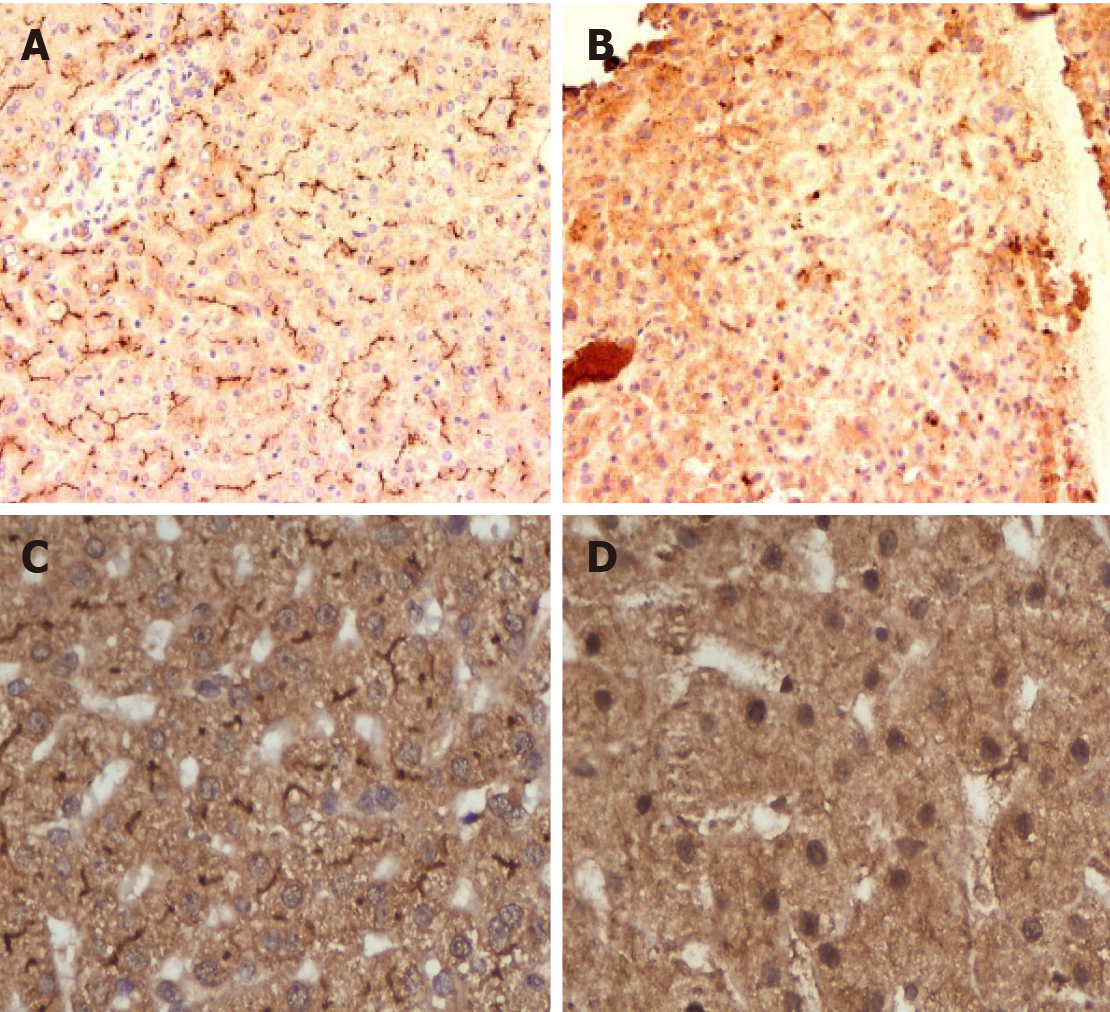
BSEP deficiency is the most common subtype of PFIC (with an estimated incidence of 1 in 50000 to 1 in 100000)[8]. It accounts for 37.5%-90.9% of cholestatic patients in the 9 studies that were analyzed in a systematic review[2]. Symptoms appear in the first month of life in 44% and by 3 mo of age in 72%[12]. Some patients with BSEP deficiency also present with early signs of vitamin D deficiency (rickets 3%-22%), vitamin K deficiency (bleeding 8%) or cholelithiasis (28%)[8,12]. High serum alanine aminotransferase and alpha-fetoprotein levels, early liver failure, cholelithiasis, HCC, very low biliary BA concentration, and negative BSEP canalicular staining suggesting PFIC2 (Figures 2A and 2B)[12]. In infancy, PFIC2 has mild to severe portal and lobular fibrosis with bridging fibrosis. Beyond infancy, advanced portal and lobular fibrosis are observed[2]. Early progression to HCC is described[19]. For more details see Table 1.
Of the 252 patients with cholestatic liver disease screened for ABCB11, 88 (34.9%) were identified with at least one disease-causing BSEP mutation with the median age of disease onset being 0.75 years[16]. 64.3% had no disease-causing mutation but at least one BSEP SNP. Amongst the common SNPs, p.A1028A was found in 197 of 252 families and p.V444A in 204 of 252 families of this cohort. The most common PFIC-2 mutations were p.E297G, p.D482G, and p.N591S. Apart from PFIC2, ABCB11 mutations cause BRIC2, ICP, contraceptive induced cholestasis and drug-induced cholestasis[16]. Four potential ABCB11 mutations and a donor splice site mutation (intron 19) were identified in 147 women with ICP[17]. Please also see the section on BRIC2 and ICP.
A global consortium on BSEP deficiency categorized the mutations as BSEP1 [p.D482G (c.1445A>G) or p.E297G (c.890A>G) mutation], BSEP2 (at least 1 missense mutation, not p.D482G or p.E297G) or BSEP3 (mutations leading to a predicted non-functional protein)[20]. Patients with BSEP1 genotype (p.D482G and p.E297G) have residual BSEP functionality and thus have a milder phenotype as compared to patients exhibiting BSEP2/BSEP3 genotype. There is wide geographical variations in genotype with the commonest mutations found in a study from Shanghai being c.145C>T (p.Gln49Ter) and c.2594C>T (p.Ala865Val)[21]. Severe phenotypes are often associated with mutations leading to premature protein truncation or failure of protein production. Insertion, deletion, nonsense and splicing mutations exhibit little or no detectable BSEP at the hepatocyte canalicular membrane.
PFIC2 is likely to have more severe lobular injury, portal fibrosis and inflammation than PFIC1. Hepatocellular necrosis and giant cell transformation are also much more pronounced in PFIC2 than in PFIC1 and may persist with time. Majority of homozygous or compound heterozygous mutations present as severe disease in infancy with progressive liver disease which requires LT. Despite treatment, 76%-100% of PFIC2 continue to have pruritus with progression to severe liver disease and this may occur as early as 7 mo of age. Advanced portal and lobular fibrosis are seen in children beyond infancy[12]. Among patients undergoing LT, liver failure and/or HCC were detectable in about 60%. Liver samples obtained from 10 children with HCC aged 13-52 mo showed that 9 had evidence of BSEP deficiency and/or mutations in ABCB11. Recently, HCC and/or cholangiocarcinoma was described in 15% of those with PFIC2. Hence, close monitoring of these children particularly those carrying 2 null mutations is essential[22]. Children with transient neonatal cholestasis harbour non-null variants, express immune-histochemically detectable BSEP and have better outcomes[21].
The global NAPPED consortium reported that 23.1% PFIC2 had undergone SBD and 46.1% had undergone LT[20]. In total, 16 (6%) [BSEP1: n = 3/72 (4%), BSEP2: n = 8/136 (6%), BSEP3: n = 5/56 (9%)] died prior to LT at a mean age of 1.6 years. NLS was 32% at 18 years of age. Five patients underwent LT beyond 18 years of age (19.6-27.5 years). Patients with mutations categorized as BSEP1 had better long-term outcomes than those with BSEP2 or BSEP3, with a median NLS of 20.4 years, 7 years and 3.5 years respectively. The incidence of HCC increased with the genotype severity from 4% in BSEP1 to 7% in BSEP2 and 34% in BSEP3. HCC occurred in 3% of patients who had SBD as against 7% who had not received SBD[20].
This autosomal recessive condition is caused by mutations of the MDR3 glycoprotein, which is coded by the ABCB4 gene located on chromosome 7q21. The gene consists of 27 coding exons spanning-74 kb[23]. ABCB4/MDR3 P-glycoprotein consists of six intracellular domains and six extracellular loops separated by twelve transmembrane domains. The protein contains two intracytoplasmic ATP-binding domains, also known as nucleotide-binding domain. The nucleotide-binding domains provide energy for the trans-membrane transfer of the substrate against the concentration gradient. The trans-membrane domains in turn provide specificity for the substrate. A study on fetal liver demonstrated that ABCB4 mRNA levels are 16-fold lower compared to the normal adult liver with only faint and focal canalicular MDR3 immunostaining suggesting that ABCB4 normally develops late in gestation or possibly in the postnatal period. The protein is present in the canalicular membrane of hepatocytes and transports phosphatidyl-choline from the hepatocytes to the bile canaliculi. Phosphatidyl-choline is a key component of micelles which keeps the cholesterol in soluble form thus protecting the cholangiocytes from damage. In presence of abnormal MDR3, phosphatidyl-choline is not transported across to the bile canaliculi leading to abnormal micelle formation. Inadequate micellar formation results in presence of insoluble bile salts and cholesterol in the biliary canaliculi which damage the cholangiocytes. Cholesterol is more likely to crystallize into stones, damaging liver structures by obstructing small bile ducts.
These patients present with a wide spectrum of manifestations ranging from transient neonatal cholestasis, episodic cholestasis, gall stones, cirrhosis to end-stage liver disease[24,25]. The patients with PFIC3 have elevated GGT and alkaline phosphatase. Bile salts and cholesterol values can be normal while biliary phospholipids are significantly reduced. Age of the onset of PFIC3 can vary widely from the neonatal period to adulthood. PFIC3 often presents later as compared to PFIC1 and PFIC2, as late as adulthood in some cases[25]. The early-onset disease can present with jaundice, pruritus, variceal bleeding (portal hypertension), stunted growth, reduced bone density and learning disabilities[25]. The complete absence of canalicular staining of MDR3 protein is associated with mutations leading to a truncated protein form (Figures 2C and 2D). Those with missense mutations have faint or normal MDR3 canalicular expression. The abnormal MDR3 canalicular expression combined with low levels of biliary phospholipids is highly suggestive of MDR3 deficiency. Histopathological findings associated with PFIC3 include primarily portal-based inflammatory infiltrate with marked periportal ductular reaction. They usually progress to biliary-type cirrhosis, lobular disarray, and hepato-canalicular cholestasis.
Degiorgio et al[26] analyzed ABCB4 gene mutations in 68 PFIC3 patients and found 29 mutations in the coding region and 10 in the transmembrane domains which involved phosphatidyl-choline translocation. Delaunay et al[27] devised a classification system for the various forms of these mutations: Class I (nonsense mutations that result in a defective synthesis), class II (missense variations that prevent protein maturation), class III (missense mutants resulting in defective protein activity), class IV (unstable variations) and class V mutants (unknown pathogenicity). These classifications are useful in determining potential therapies for patients. Approximately 300 disease-causing ABCB4 variants have been reported, typically in those with homozygous status who have a rapidly progressive cholestatic liver disease with potentially life-threatening complications. Those with heterozygous status have less severe manifestations.
Patients with residual phosphatidyl-choline secretion and MDR3 expression (espe
BRIC is an autosomal recessive inherited disorder characterized by the intermittent occurrence of severe and recurrent cholestasis[29]. BRIC typically appears later than PFIC and is not progressive. Two genetic forms of BRIC are known; BRIC1 is characterized by the mutations of the ATP8B1 gene while BRIC2 presents a mutation in the ABCB11 gene. BRIC shows a non-progressive course since the protein function is only partially impaired[29]. Most of them have missense mutations located in less conserved regions of the gene. The global NAPPED consortium reported 11 BRIC2 who later presented with persistent cholestasis and progressive liver disease with pathological genetic variants[20].
ICP is the occurrence of intense pruritus in a pregnant woman without pre-existent liver disorder; beginning usually in the third trimester and resolving completely after delivery. Elevated levels of BA, aminotransferase and alkaline phosphatase are typically present. Although the disease is benign in the mothers, poor fetal outcomes have been reported. Heterozygous mutations in ABCB4, ABCB11, ATP8B1, ABCC2 and tight junction protein 2 have been associated with ICP[30]. While GGT is normal in ABCB11-related forms of estrogen-associated cholestasis, increased GGT levels could serve as a discriminating feature of ABCB4 deficiency.
The associations of heterozygous missense and nonsense ABCB4 mutations in cryptogenic liver fibrosis and biliary cirrhosis have been reported[28,31]. The his
MDR3 deficiency also leads to gallbladder disease or low phospholipid-associated cholelithiasis (GBD-1/LPAC)[32]. There is more evidence that either biallelic or monoallelic ABCB4 defects may cause or predispose patients to a wide spectrum of human liver diseases such as LPAC, small duct sclerosing cholangitis and adult biliary fibrosis or cirrhosis[28]. Liver cirrhosis accompanied by vanishing bile duct syndrome (ductopenia) in ABCB4 deficient patients have been reported[33].
ABCB11 and ABCB4 deficiency may predispose to the cholestatic liver injury induced by oral contraceptives, psychotropic drugs, selected chemotherapy drugs, statins, and antibiotics[34]. The common variant p.Val444Ala in ABCB11 is associated with an increased risk of drug-induced liver injury[35]. Most cases improve on withdrawing the drug and adding UDCA. Recent studies have demonstrated, that ABCB4 mutations (c.504C>T and c.485T>Ain exon 6 and c.2793 frameshift mutation in exon 23) are possibly involved in the development of parenteral nutrition-associated liver disease in premature infants[36].
The holistic management of patients with PFIC includes their nutritional rehabilitation, fat-soluble vitamin supplementation, control of pruritus, prevention of fibrosis progression and LT for those with end-stage liver disease.
Chronic malnutrition due to cholestasis and steatorrhoea affects almost all children with PFIC, especially those with PFIC1[37,38]. Apart from cholestasis-related malabsorption, complex interactions of the liver with the hypothalamus-pituitary-adrenal axis in chronic cholestasis have been advocated as one of the mechanisms for growth failure in these children[39]. Severe cholestasis leads to deficiency of fat-soluble vitamins (FSV) with decreased bone mineral density, often refractory to FSV supplementation[38]. Meticulous nutritional assessment should be performed and documented at the first visit. The goal of nutritional rehabilitation should be to give calories around 125% of the recommended daily dietary allowance for the ideal weight targeting around 180-200 cal/kg/d along with high protein about 2-3 g/kg/d[38]. FSV should be supplemented orally: Vitamin A 5000-10000 IU/d; Vitamin D (cholecalciferol) 2000-5000 IU/d; Vitamin E 50-400 IU/d and Vitamin K 5 mg/wk to 5 mg/d[40]. Weekly or daily supplementation of FSV, especially vitamin D, is more effective than large bolus doses for the treatment of vitamin D deficiency in children with chronic liver disease[41]. Refractory deficiency or those with significant bony changes should be treated with calcitriol at a dose of 0.05-0.2 μg/kg[40]. The oral water-soluble liquid formulation of FSV containing vitamins A, D, E and K have shown to be more efficacious than conventional preparation in infants and children with cholestatic liver disease. Medium-chain triglycerides should be supplemented as they are absorbed directly into the portal circulation and are not affected by cholestasis[40].
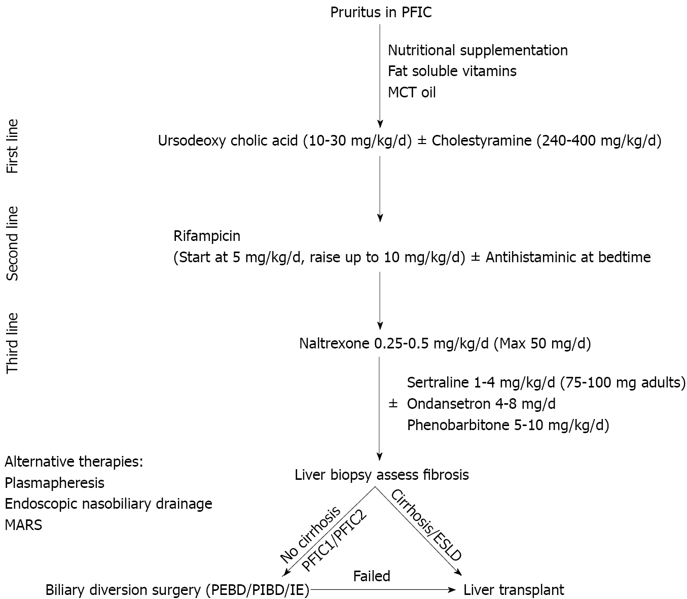
Pruritus is the most disabling symptom of PFIC disturbing daily activities, schooling and sleep. A step-up approach of medical therapy is advocated in children with pruritus. SBD is a very useful tool in medical non-responders (Figure 3). Table 2 lists the drugs used for the management of pruritus with their mechanism of action, dose and adverse effects. Apart from medical therapy, trimmed nails, full-sleeve clothing-stockings during sleep and well moisturized skin are advocated. Various tools like visual analog scale, Whitington scale or 5D-pruritus score have been used for objective measurement of pruritus and to assess response to medications or surgical therapy[42].
| Drug | Mechanism of action | Dose | Adverse effects |
| Ursodeoxycholic acid | Protection of cholangiocytes from the hydrophobic bile acids; Choleretic action through both bile acid dependent (cholehepatic shunt) and independent pathway; Protection of hepatocytes from bile acid induced apoptosis; Direct membrane stabilizing effect in cholangiocytes; Up-regulate synthesis, apical insertion & activation of BSEP & Mrp2 via Ca2+ and PKC-dependent mechanisms or via activation of p38 MAPK and Erk-1/2–dependent mechanisms in animal models | 10-30 mg/kg/d | Adverse effects rare: Severe vomiting or diarrhoea |
| Rifampicin | Activates pregnane X receptor leading to decrease in autotoxin level thus leading to decrease in lysophosphatidic acid synthesis and down-regulation of TRP vanilloid 1; Upregulates multidrug-resistance protein 2; Activates enzymes UDP-glucuronosyltransferase-1A and cytochrome P450-3A4 and stimulates 6α-hydroxylation of bile acids, promoting urinary excretion of dihydroxy and monohydroxy bile acids | 5-10 mg/kg/d | Adverse effects rare: Hepatotoxicity, vomiting |
| Bile acid sequestrants: Cholestyramine, colestipol, colesevelam | Non-absorbable anion exchange resins that bind bile acids, cholesterols and other compounds in the intestinal lumen and prevent their enterohepatic circulation | 240-500 mg/kg/d; Usually administered mixed with juice | Palatability, steatorrhoea, constipation, intestinal obstruction from inspissations, hyperchloremic metabolic acidosis; Growth failure; Decreased absorption of other drugs (e.g., UDCA) if not spaced; Need to be spaced from food |
| Opioid antagonists: Naltrexone | Reduces central opioidergic tone, believed to be raised in patients with cholestatic pruritus; Decreasing plasma levels of endogenous opioids like enkephalins | Gradually increment starting at 12.5 mg/d increasing every 3-7 d till pruritus reduces | Opioid withdrawal-like symptoms including abdominal pain, tachycardia and hypertension |
| Selective serotonin reuptake inhibitors: Sertraline | Exact mechanism of action not elucidated; Mediates its effect through serotonergic signals in the central nervous system that provide inhibitory signals to the itch pathways; Neuropharmacologic inhibition of stress | Adults: 75-100 mg/d; Children: 2.2 mg/kg/d | Adverse effects: Allergic reaction, behavioural issues, diarrhoea, insomnia, dizziness, high first pass metabolism-risk of hepatotoxicity |
UDCA leads to complete resolution of pruritus in about a third of children with PFIC2[38] and response is mainly dependent on the mutation and resultant residual protein expression[24]. Long-term therapy with UDCA has been shown to reverse fibrosis in MDR3 heterozygotes with residual protein expression[43]. The role of rifampicin in management of pruritus has been established by meta-analyses (pooled odds ratio of 15.2 for complete/partial relief) as compared to placebo/alternative therapy[44]. Bile acid sequestrants are less favourable in children due to non-palatability[38]. Spacing is needed from food and other drugs leading to loss of compliance and optimal effect[45]. Opioid antagonists can ameliorate pruritus, although with increased incidence of adverse events[44]. Sertraline, a selective serotonin reuptake inhibitor is also beneficial for the relief of pruritus. Sertraline has high first-pass metabolism in the liver and should be used with caution. The other drugs with equivocal efficacy for control of pruritus in PFIC include ondansetron, phenobarbitone and antihistaminics[40,41]. Newer modalities to treat pruritus of cholestasis that have not been extensively analyzed in studies include albumin dialysis using molecular adsorbent recirculating system, plasmapheresis and endoscopic nasobiliarydrainage[46]. Plasmapheresis reduces autotaxin activity thereby reducing lysophosphatidic acid and reducing pruritus in ICP.
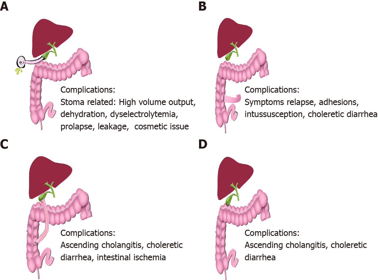
Commonly used SBD include partial external biliary diversion (PEBD), ileal exclusion (IE) and partial internal biliary diversion (PIBD). Figure 4 shows a schematic representation of the various diversion surgeries, techniques and their common complications. PEBD, although being the most widely used diversion surgery is associated with a host of stoma-related complications[47-49]. Recently, external fistulae have been intubated with gastrostomy devices for intermittent drainage[49]. IE[50,51] and PIBD[52,53] are therapeutic options in those with failed PEBD or for better cosmetic results, especially in adolescents. Recent studies on SBD with their outcome and complications have been tabulated in Table 3[51-55]. Biliary diversion leads to a relatively higher ratio of cholic acid to CDCA in bile, reduced taurine-to-glycine conjugate ratio, increase in secondary bile acid pool, ultimately causing bile to become more hydrophilic[55]. Total biliary diversion involves diverting all the bile flow from the common bile duct to the exterior through a jejunal loop. It is usually reserved for pruritus refractory to PEBD or to prevent and treat allograft steatosis in post LT PFIC1[56]. Irrespective of the technique used, biliary diversion surgeries lead to a decrease in the concentration of BA, improvement in pruritus score, growth spurt, histological improvement and even reversal of fibrosis in some cases[38,47]. There are no head-to-head trials to compare the efficacy and safety of these techniques. Post SBD, the child should be monitored for improvement in pruritus, changes in growth parameters and for decrease in BA and bilirubin in the serum. Recently, the NAPPED consortium and another meta-analysis have shown improved NLS in those with common missense mutations (D482G or E297G) and in those with more than 75% reduction in BA post SBD[20,57]. Around 18%-39% of children undergoing SBD require LT in 5-10 years of follow-up, mostly those with advanced fibrosis at the time of SBD and those with failure of SBD[51-55]. Those with advanced fibrosis do not show a reduction in serum BA with biliary diversion surgeries, hence a liver biopsy should precede the surgery[57]. The choice of biliary diversion technique depends on the patient’s preference and experience and comfort of the surgeon/centre. Health-related quality of life is comparable between those undergoing PEBD for PFIC and healthy controls.
| Study | Type of biliary diversion | No of patients | Median follow up | Outcome | Adverse events |
| Yang et al[54] (2009) | PEBD | 14 (11-PFIC) | 3.1 yr (2-5.7) | Resolution of pruritus: 50%; Decrease in pruritus: 21%; Decrease in serum bile acids; Improvement in growth; Improved quality of life; No response in 2 patients with advanced fibrosis; 21.4% were listed for LT at mean follow-up 3.2 yr (all had advanced fibrosis pre-PEBD) | 3 developed stoma prolapse; Post-op bleed and wound dehiscence in 1 each |
| Schukfeh et al[55] (2012) | PEBD | 24 | 9.8 yr (1.6-14.3) | Resolution of pruritus with normalization of bile acids in 54%; 37.5% received LT at mean 1.9 yr; All of them had failed PEBD & 78% of them had cirrhosis pre-PEBD | Stomal prolapse in 2; Cholangitis, dyselectrolytemia, GI bleed and intestinal obstruction in 1 each |
| Wang et al[50] (2017) | PEBD; IE; PIBD | 39; 11; 7; (38 PFIC & 20 alagillesyndrome) | 24 mopostsurgery | Decrease in severe pruritus-54% in PFIC1 and 30% in PFIC2; Trend towards decreased pruritus after IE and PIBD; PEBD but not IE led to decrease in bilirubin and ALT in PFIC1; 23.7% of PFIC underwent LT post diversion | PEBD: Dehydration/dyselectrolytemia in 4; Stoma prolapse in 3; Intestinal ischemia & bowel obstruction in 1 each; IE; Dyselectrolytemia-2; PIBD: Dyselectrolytemia in 2, intestinal ischemia & intussusception-1 each |
| Cielecka-Kuszyk et al[47] (2019) | PEBD | 4 (all PFIC2) | > 10 yr | Resolved cholestasis in 3; Reversal of fibrosis in 2 | |
| Bull et al[48](2018) | PEBD; IE | 57; 6 | Sustained improvement in pruritus: PFIC1-57%; PFIC2 (D482G/E297G mutations)-76%; PFIC2 other mutations-33%; Improvement in bilirubin and bile acids; Improvement in growth; 27% of PFIC1 & 31% of PFIC2 were listed or received LT (less often in D482G/E297G) | Dehydration/dyselectrolytemia due to high stoma output seen in 6 patients (1 died); Cholangitis in 3; Bile stagnation in 2; Stoma bleed in 1 | |
| Van Wessel et al[20] (2020) | PEBD; IE; PIBD | 47; 13; 1; (all PFIC2) | 8.4 yr (1.6-12) | Relief in pruritus – sustained: 54%; Transient: 17%; None: 29%; Relief in pruritus more common in BSEP1 mutations (66%) vs BSEP2 (36%) & BSEP3 (0%); Decrease in serum bile acids, bilirubin, AST & ALT; A 75% reduction in bile acids or decrease to a level < 102 µmol/L post diversion predicts long term NLS of > 15 yr; Biliary diversion associated with higher NLS: HR 0.51; 95%CI: 0.29-0.91, P = 0.02 | |
| Bjørnland et al[49] (2021) | PEBD | 33; (25 PFIC) | 10 yr (0.6-25.2) | Decrease in bile acids 1 wk post-op predictive of successful drainage; 39% received LT or were listed LT at a median follow up of 10 yr | 42% early post op complications; Long term stoma related complications in 55%-20% secondary surgeries |
| Van Vaisberg et al[51] (2020) | IE | 11 | 5 yr | Significant relief in pruritus: 8 (72.7%); 2/11 (18.2%) progressed to ESLD within a year and were listed for LT | Intussusception in 1; No diarrhoea |
| Foroutan et al[53] (2020) | PIBD | 44 | 54 mo (10-105) | Significant decrease in jaundice and pruritus | Ascending cholangitis in 19.2%; No difference in cholangitis between standard procedure and PIBD with anti-reflux valve |
| Chen et al[52](2018) | PIBD | 34; (PFIC1-10, PFIC2-14, PFIC3-5) | - | Decreased bilirubin and bile acids; Improved growth; 2 (5.9%) underwent LT at 20 & 39 mo post PIBD | Dyselectrolytemia/dehydration in 2; Relapse of symptoms in 4 |
| Agarwal et al[38] (2016) | PIBD; PEBD | 3; 1 | 2 yr (1-2) | PIBD: Decrease in pruritus score, improved growth & decreased serum bile acids; PEBD: Failed; One with failed PEBD needed LT in 7 yr; Rest all survived with native liver at mean follow up 8 yr | No complications with PIBD |
| Bull et al[48](2018) | PEBD; IE | 57; 6 | Sustained improvement in pruritus: PFIC1-57%; PFIC2 (D482G/E297G mutations)-76%; PFIC2 other mutations-33%; Improvement in bilirubin and bile acids; Improvement in growth; 27% of PFIC1 & 31% of PFIC2 were listed or received LT (less often in D482G/E297G) | Dehydration/dyselectrolytemia due to high stoma output seen in 6 patients (1 died); Cholangitis in 3; Bile stagnation in 2; Stoma bleed in 1 |
LT is indicated in children with end-stage liver disease, advanced fibrosis and those with refractory pruritus despite biliary diversion surgeries, yet it is fraught with its unique complications and conundrums in children with PFIC[58]. PFIC is one of the most common indications accounting for 13.4% of total pediatric LT[59]. LT in patients with PFIC2 and PFIC3, where the mutation affects only the liver has resulted in excellent graft and patient survival[37]. On the other hand, due to a wide extrahepatic tissue distribution of FIC1 expression, LT for PFIC1 has been associated with a high risk of complications due to the involvement of these extrahepatic sites after LT[60]. There are three unique issues related to LT in PFIC: (1) High rate of graft failure, allograft steatosis and diarrhea in PFIC1; (2) Antibody-induced BSEP deficiency leading to graft loss in PFIC2; and (3) Utilization of heterozygous family members as donors in living donor LT program.
Allograft steatosis, refractory diarrhea and graft failure in PFIC1: Post LT in PFIC1, the graft liver starts to secrete and excrete normal amounts of bile salts which can’t be handled by the intestines as the intestinal FIC1 is still defective leading to intractable diarrhea[60]. This plausible mechanism for intractable diarrhea is further strengthened by the clinical response to bile adsorption resins and total biliary diversion. FIC1 in the intestine which has a strong correlation to SLC10A2, the ileal sodium/bile acid co-transporter is defective in PFIC1 leading to the impaired enterohepatic circulation of bile salts[61]. When the intestine of these patients which are defective in FIC1/SLC10A2 starts receiving a normal amount of BA secreted by the graft liver, it leads to bile acid diarrhea, steatosis and graft fibrosis necessitating re-transplant[60]. Hori et al[37] reported chronic diarrhea in 91%, grade 3 steatosis in 72.7% and significant graft fibrosis in 82%; 27% died at a median of 12 years post LT and 3 required re-transplant. Davit-Spraul et al[25] reported extrahepatic complications post LT in 100% of PFIC1 and one-third required re-transplant for massive steatosis. There is a recurrence of steatosis and graft failure even in the second graft[12,60]. The genetic analysis predicts the patients likely to develop these complications and thus pre-LT genetic analysis should be considered mandatory for patients with PFIC1[60]. Total internal biliary diversion during the time of LT has been described as a modality by the Kasahara group to prevent complications of diarrhea and steatosis post LT[62].
Antibody induced BSEP deficiency: Post-transplant recurrence of cholestasis in PFIC2 has been described due to de novo polyclonal antibodies directed against extracellular loop 1 domain of BSEP protein in the sera and on the canalicular membrane[63]. Patients with severe mutations with resultant complete absence of BSEP are typically prone to this complication as the BSEP of the transplanted liver is recognized as a foreign new antigen by the recipient’s immune system, usually seen after an episode of acute rejection. Initial treatment of antibody-induced BSEP-deficiency disease is increased immunosuppression followed by either rituximab, bortezomib, intravenous immunoglobulin or plasmapheresis[64]. Those refractory to the above treatment may be considered for allogeneic hematopoietic stem cell transplantation[65]. Re-transplant may be considered in those refractory to all therapy but the threat of recurrence in the re-transplanted liver is high[63].
Donor issues in living-related liver transplant: There have been apprehensions about using family members as donors, who may be heterozygous. Bassas et al[66] described living-related donor LT in 13 children with PFIC with good outcomes and no recurrence of symptoms in the recipients. Other studies have also reported favourable outcomes in PFIC2 and PFIC3 with living-related LT[67]. Genetically proven heterozygous family members have been used as donors without recurrence of symptoms in the recipients. Currently, there is not enough evidence to perform a genetic analysis of the related donor before LT. It remains to be seen if the heterozygous donor liver in PFIC3 patients may predispose the recipient to drug-induced cholestasis, contraceptive induced cholestasis, ICP or GBD-1/LPAC.
Gene therapy: The additional complications associated with LT warrant the consideration of innovative therapeutic modalities like gene therapy. Recent approval for Adenovirus Associated viral (AAV) vector-based gene therapy for other monogenic disorders, raises hope for such future therapies for PFIC[68]. The liver tropism of AAV vectors is a major advantage in the treatment of PFIC as this would imply the need for a lower vector dose for therapeutic efficacy. Higher AAV vector doses have been shown to cause adverse effects ranging from transient liver inflammation to even progressive liver failure and deaths in few reports[69]. Gene therapy can be non-integrating where each cell division post therapy will lead to some loss of episomal AAV vector genome or integrating in vivo where these integrated genes are copied during cell division and passed on to the daughter cells without any loss of vector genome upon cell division[68]. The benefit of integrating gene therapy is that a limited number of corrected hepatocytes will repopulate the liver and correct the defect[68,70]. Theoretically, in vivo non-integrating AAV mediated gene therapy can be successful in PFIC3 as establishing ABCB4 expression in a proportion of hepatocytes. This can partially correct phosphatidylcholine transport and prevent the progression of the disease. Gene therapy for PFIC has only been done successfully in double knockout murine models (Abcb4-/-) to date[71]. Treatment with modified mRNA variants encoding human ABCB4 encapsulated in lipid nanoparticles lead to de novo expression of functional ABCB4 and restored phospholipid transport in cultured cells as well as Abcb4-/-mice. Hepatocyte damage due to accumulated BA in PFIC1 and 2; and decreased membrane stability in PFIC1 would warrant the correction of the genetic defect in all the hepatocytes, thus needing an integrating gene therapy strategy[68]. Tumor genes is a possible major risk factor with integrating gene therapy as seen in trials on patients with severe combined immunodeficiency[72]. The evolution of CRISPR/Cas9 based gene-editing tools that exhibit fewer off-target effects raises hope for integrating in vivo controlled gene therapy in future[73].
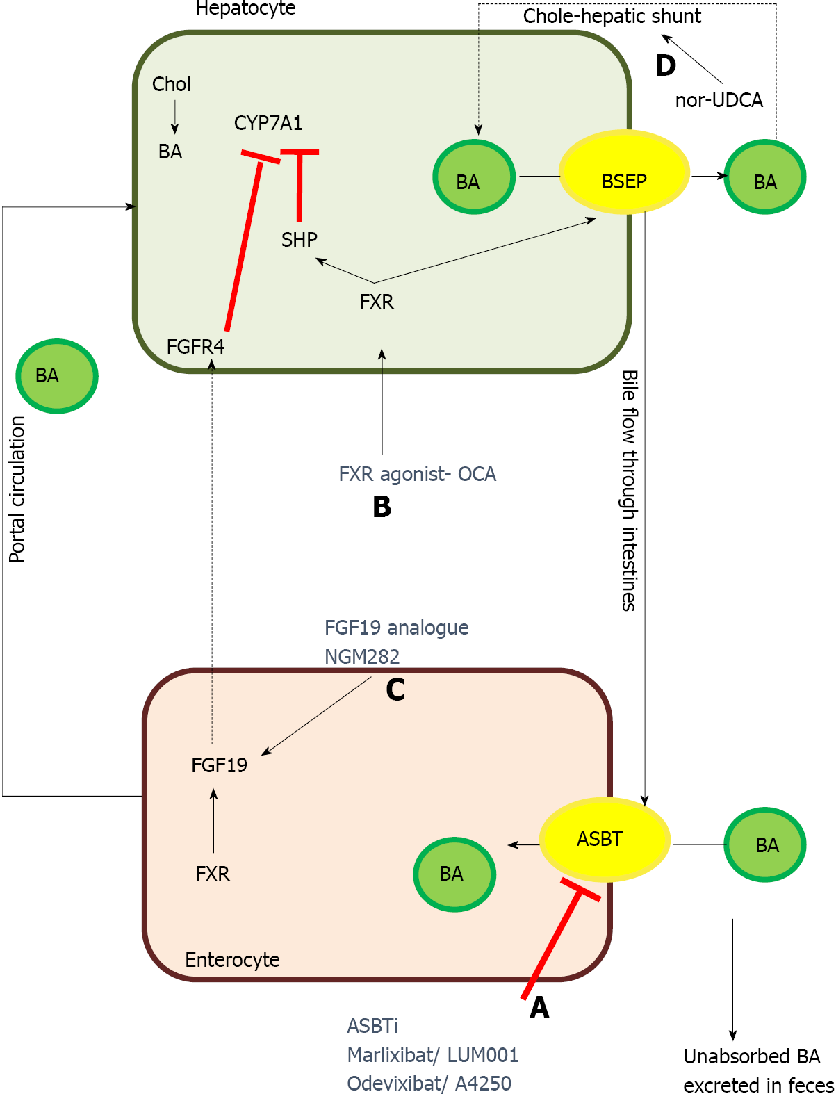
Newer targets for therapy in PFIC: Several drugs and therapeutic molecules are currently being investigated for their therapeutic efficacy and safety in PFIC1 and 2. Table 4 and Figure 5 describes the various experimental drugs being evaluated for their therapeutic role in PFIC. Among them, the class of drugs that have generated the maximum interest are those which inhibit the ileal ASBTs.
| Drug | Mechanism of action | Clinical trials and current status | Notes |
| Maralixibat/LUM001 | Apical sodium-dependent bile acid transporter inhibitor | NCT04185363: Open label phase III trial; Recruiting patients; NCT03905330: MARCH-PFIC trial; Randomized controlled trial, recruiting patients; NCT04729751: RISE trial in infants; Open label phase II safety study; NCT04168385: Long term safety study; NCT02057718; Open label phase II trial; Completed | Orphan drug designation by FDA; Breakthrough therapy for PFIC2 |
| Odevixibat/A4250 | Selective inhibitor of ileal bile acid transporter | NCT03566238: PEDFIC 1 study; Phase III, open label, randomized controlled trial; Ongoing; NCT04483531: Expanded access study including patients not enrolled in PEDFIC 1 study | Orphan drug designation by FDA; Fast track designation for PFIC |
| 4-PB/GPA | Prolongs degradation rate & increases cell surface expression of BSEP & functions as a chemical chaperone to correct the misfolded proteins | Leads to long term reduction in serum BA, improvement in liver biochemistry as well as relief of pruritus; Increased canalicular localization of E297G and D482G BSEP mutants; GPA more palatable, has lower sodium, doesn’t interact with rifampicin; Doses: 4-PB: 500 mg/kg/d; GPA: 8 g/m2/d | 4-PB FDA approved for urea cycle defect |
| Ivacaftor | Rescues the function of missense mutations in the nucleotide binding domains of BSEP & MDR3 | In vitro correction of binding domain missense mutation (T463I) of BSEP; Improved phospholipid secretion activity in mutant ABCB4 | In vitro studies; Animal studies |
| Oxcarbazepine | Nerve stabilizing effect; Enzyme inducer – possible role in potentiating action of 4-PB | Single case report on its combined use with 4-PB and maralixibat | |
| Gentamicin | Induce readthrough in nonsense mutation | In vitro increased readthrough in 6 common nonsense mutation of BSEP leading to increased canalicular expression of bile salt transporter | |
| FXR agonist (Obeticholic acid) | Farsenoid X receptor agonist | No trials in PFIC; Safe and efficacious in treatment of PSC and non-alcoholic steatohepatitis | FDA approved for PSC |
| Nor-UDCA | Side-chain-shortened derivative of UDCA; Increases cholehepatic shunt | No trials in PFIC; NCT03872921: Ongoing phase III randomized controlled trial in PSC | |
| Steroids | Possible upregulation of BSEP transporter? Up-regulation of sodium taurocholate copeptide transporterproviding increased gradient for BSEP | Only case reports and animal studies | |
| NGM282 | FGF19 analogue | NGM282 inhibited bile acid synthesis and decreased fibrosis markers, without change in alkaline phosphatise level | |
| Bezafibrate | Peroxisome proliferator activated receptor agonist | Bezafibrate reduced pruritus and cholestasis in 2 out of 3 children with PFIC1 and improved lipid profile in all |
Apical sodium bile acid transporter inhibitors: ASBTi is currently under various stages of clinical trials include maralixibat and odevixibat (A4250). Currently, there are 4 registered clinical trials for maralixibat including one in infants and two clinical trials for odevixibat. The ASBTi interfere with bile salts reabsorption in the terminal ileum and hence greatly reduce the enterohepatic circulation of bile salts which are then excreted in the stool. ASBTi thus shrinks the bile acid pool and helps reduce BA and in turn improves cholestasis[74]. Odevixibat is as effective as PEBD in reducing BA as well as pruritus in a child with PFIC2 thus raising hope of a very effective medical management of cholestasis[75]. However, on stopping therapy after the completion of the trial, the patient had a relapse of symptoms and underwent PEBD. The preliminary results from the trials of maralixibat and odevixibat have shown that these ASBTi reduce BA and pruritus while improving growth[74,75].
Ivacaftor: Ivacaftor (VX-770), a CFTR/ABCC7 potentiator has been demonstrated to cause functional rescue of missense mutations of ABCB11 and ABCB4 located in a highly conserved ABC transporter motif[76]. In vitro molecular modelling showed that ivacaftor led to the functional rescue of mutant ABCB4 resulting in increased phos
4 phenyl butyrate: 4-phenyl butyrate (4-PB), an FDA approved drug for urea cycle defect acts by 2 mechanisms to correct the defect in BSEP missense mutations: (1) It modulates the short-chain ubiquitination to prolong the degradation rate of cell surface resident BSEP and hence increases the cell surface expression of BSEP; and (2) Functions as a chemical chaperone to correct the misfolding of some endoplasmic reticulum-retained BSEP missense mutants[77]. In vitro studies in HEK293 and MDCKII cell lines showed increased cell surface expression of wild-type E297G and D482G BSEP. This resulted in an increased taurocholic acid transport on treatment with 4-PB, long term reduction in BA, improved presence of BSEP at canalicular membrane and relief of pruritus. 4-PB therapy was effective at a dose of 500 mg/kg/d with no adverse events but with relapse of symptoms after stopping therapy[78]. However, concerns have been raised about the palatability of this drug, compliance and hepatotoxicity potential when combined with rifampicin. Glycerol phenylbutyrate was shown to be equally efficacious with good tolerance and adherence. Malatack et al[79] described a case report of a PFIC2 with a missense mutation in BSEP who improved with combination therapy of 4-PB, maralixibat and oxcarbazepine. They hypothesized that enzyme inducer action of oxcarbazepine augments conversion of 4-PB to phenylacetate leading to synergistic effect apart from its usual nerve stabilizing effect[79]. Although 4-PB is safe in most studies, a recent report of bipolar disorder in an adolescent on 4-PB raises concerns[80].
Readthrough therapy-gentamicin: Gentamicin has been shown to induce readthrough leading to the expression of a full-length protein in stable MDCK clones. Gentamicin in combination with 4-PB significantly increased the readthrough level of common nonsense mutation studied (p.R415X, p.R470X, p.R1057X, p.R1090X) in HEK293 cells. The maximum response was seen for the p.R1090X mutation with a 40% increase in taurocholate transport and correct localization at the cell membrane[81]. There has been an emphasis on mutation-specific therapies with: (1) Gentamicin and ataluren likely to suppress nonsense mutations by promoting the readthrough of premature stop codons; and (2) U1-small nuclear-RNA likely to rescue splicing for several ATP8B1 mutations located at donor, acceptor and splice sites[81].
Other drugs: Nor-UDCA is a side-chain shortened derivative of UDCA with relative resistance to amidation thus increasing the cholehepatic shunt and in turn increases the bile salt-dependent bile flow[82]. Peroxisome proliferator-activated receptor agonist bezafibrate has been reported to correct dyslipidemia and reduce pruritus and cholestasis in children with PFIC1 and other diseases[83]. Rapamycin restores bile acid excretion, attenuates hepatocyte damage and extends the lifespan of Abcb11b mutant zebrafish which is the ortholog of human ABCB11. Other drug categories which could theoretically be beneficial but have not yet been tried in clinical trials include FXR agonist and fibroblast growth factor-19 analogue[84]. Obeticholic acid is a very potent FXR agonist, promotes transcription of BSEP, improves histopathology and reverse fibrosis and could therefore be useful in patients with PFIC2[84].
To conclude, there has been a recent emphasis on understanding the genetic spectrum of PFIC, their phenotype, natural course and long-term outcome. Genotype correlates well with phenotype in PFIC2 but not in PFIC1. The expanded role of the heterozygous transporter defects presenting late needs to be suspected and identified even in adulthood. Medical therapy and SBD form the cornerstone of the management of pruritus. LT in these children is associated with unique issues like a high rate of intractable diarrhea, growth failure, steatohepatitis and graft failure in PFIC1 and antibody-mediated BSEP deficiency. There is a promising role of ASBT inhibitors in the management of cholestasis.
Provenance and peer review: Invited article; Externally peer reviewed.
Peer-review model: Single blind
Specialty type: Gastroenterology and hepatology
Country/Territory of origin: India
Peer-review report’s scientific quality classification
Grade A (Excellent): 0
Grade B (Very good): B
Grade C (Good): 0
Grade D (Fair): D
Grade E (Poor): 0
P-Reviewer: Cheung MCM, Dabbous H S-Editor: Wang JJ L-Editor: A P-Editor: Wang JJ
| 1. | Jacquemin E. Progressive familial intrahepatic cholestasis. Clin Res Hepatol Gastroenterol. 2012;36 Suppl 1:S26-S35. [RCA] [PubMed] [DOI] [Full Text] [Cited by in Crossref: 151] [Cited by in RCA: 154] [Article Influence: 11.8] [Reference Citation Analysis (0)] |
| 2. | Baker A, Kerkar N, Todorova L, Kamath BM, Houwen RHJ. Systematic review of progressive familial intrahepatic cholestasis. Clin Res Hepatol Gastroenterol. 2019;43:20-36. [RCA] [PubMed] [DOI] [Full Text] [Cited by in Crossref: 48] [Cited by in RCA: 90] [Article Influence: 15.0] [Reference Citation Analysis (0)] |
| 3. | Takatsu H, Tanaka G, Segawa K, Suzuki J, Nagata S, Nakayama K, Shin HW. Phospholipid flippase activities and substrate specificities of human type IV P-type ATPases localized to the plasma membrane. J Biol Chem. 2014;289:33543-33556. [RCA] [PubMed] [DOI] [Full Text] [Cited by in Crossref: 83] [Cited by in RCA: 101] [Article Influence: 9.2] [Reference Citation Analysis (0)] |
| 4. | Groen A, Romero MR, Kunne C, Hoosdally SJ, Dixon PH, Wooding C, Williamson C, Seppen J, Van den Oever K, Mok KS, Paulusma CC, Linton KJ, Oude Elferink RP. Complementary functions of the flippase ATP8B1 and the floppase ABCB4 in maintaining canalicular membrane integrity. Gastroenterology. 2011;141:1927-37.e1. [RCA] [PubMed] [DOI] [Full Text] [Cited by in Crossref: 93] [Cited by in RCA: 94] [Article Influence: 6.7] [Reference Citation Analysis (0)] |
| 5. | Strautnieks SS, Bull LN, Knisely AS, Kocoshis SA, Dahl N, Arnell H, Sokal E, Dahan K, Childs S, Ling V, Tanner MS, Kagalwalla AF, Németh A, Pawlowska J, Baker A, Mieli-Vergani G, Freimer NB, Gardiner RM, Thompson RJ. A gene encoding a liver-specific ABC transporter is mutated in progressive familial intrahepatic cholestasis. Nat Genet. 1998;20:233-238. [RCA] [PubMed] [DOI] [Full Text] [Cited by in Crossref: 766] [Cited by in RCA: 674] [Article Influence: 25.0] [Reference Citation Analysis (0)] |
| 6. | Folmer DE, Elferink RP, Paulusma CC. P4 ATPases - lipid flippases and their role in disease. BiochimBiophys Acta. 2009;1791:628-635. [RCA] [PubMed] [DOI] [Full Text] [Cited by in Crossref: 93] [Cited by in RCA: 97] [Article Influence: 6.1] [Reference Citation Analysis (0)] |
| 7. | Chen F, Ellis E, Strom SC, Shneider BL. ATPase Class I Type 8B Member 1 and protein kinase C zeta induce the expression of the canalicular bile salt export pump in human hepatocytes. Pediatr Res. 2010;67:183-187. [RCA] [PubMed] [DOI] [Full Text] [Cited by in Crossref: 23] [Cited by in RCA: 25] [Article Influence: 1.7] [Reference Citation Analysis (0)] |
| 8. | Vitale G, Gitto S, Vukotic R, Raimondi F, Andreone P. Familial intrahepatic cholestasis: New and wide perspectives. Dig Liver Dis. 2019;51:922-933. [RCA] [PubMed] [DOI] [Full Text] [Cited by in Crossref: 52] [Cited by in RCA: 47] [Article Influence: 7.8] [Reference Citation Analysis (4)] |
| 9. | Stapelbroek JM, Peters TA, van Beurden DH, Curfs JH, Joosten A, Beynon AJ, van Leeuwen BM, van der Velden LM, Bull L, Oude Elferink RP, van Zanten BA, Klomp LW, Houwen RH. ATP8B1 is essential for maintaining normal hearing. Proc Natl Acad Sci U S A. 2009;106:9709-9714. [RCA] [PubMed] [DOI] [Full Text] [Cited by in Crossref: 90] [Cited by in RCA: 98] [Article Influence: 6.1] [Reference Citation Analysis (0)] |
| 10. | Ray NB, Durairaj L, Chen BB, McVerry BJ, Ryan AJ, Donahoe M, Waltenbaugh AK, O'Donnell CP, Henderson FC, Etscheidt CA, McCoy DM, Agassandian M, Hayes-Rowan EC, Coon TA, Butler PL, Gakhar L, Mathur SN, Sieren JC, Tyurina YY, Kagan VE, McLennan G, Mallampalli RK. Dynamic regulation of cardiolipin by the lipid pump Atp8b1 determines the severity of lung injury in experimental pneumonia. Nat Med. 2010;16:1120-1127. [RCA] [PubMed] [DOI] [Full Text] [Full Text (PDF)] [Cited by in Crossref: 110] [Cited by in RCA: 120] [Article Influence: 8.0] [Reference Citation Analysis (0)] |
| 11. | Bull LN, Morotti R, Squires JE. ATP8B1 Deficiency. 2001 Oct 15. In: GeneReviews® [Internet]. Seattle (WA): University of Washington, Seattle; 1993–2021. [PubMed] |
| 12. | Al-Hussaini A, Lone K, Bashir MS, Alrashidi S, Fagih M, Alanazi A, AlYaseen S, Almayouf A, Alruwaithi M, Asery A. ATP8B1, ABCB11, and ABCB4 Genes Defects: Novel Mutations Associated with Cholestasis with Different Phenotypes and Outcomes. J Pediatr. 2021;236:113-123.e2. [RCA] [PubMed] [DOI] [Full Text] [Cited by in Crossref: 7] [Cited by in RCA: 9] [Article Influence: 2.3] [Reference Citation Analysis (1)] |
| 13. | van der Woerd WL, van Haaften-Visser DY, van de Graaf SF, Férec C, Masson E, Stapelbroek JM, Bugert P, Witt H, Houwen RH. Mutational analysis of ATP8B1 in patients with chronic pancreatitis. PLoS One. 2013;8:e80553. [RCA] [PubMed] [DOI] [Full Text] [Full Text (PDF)] [Cited by in Crossref: 4] [Cited by in RCA: 8] [Article Influence: 0.7] [Reference Citation Analysis (0)] |
| 14. | Li L, Deheragoda M, Lu Y, Gong J, Wang J. Hypothyroidism Associated with ATP8B1 Deficiency. J Pediatr. 2015;167:1334-9.e1. [RCA] [PubMed] [DOI] [Full Text] [Cited by in Crossref: 11] [Cited by in RCA: 15] [Article Influence: 1.5] [Reference Citation Analysis (0)] |
| 15. | van Wessel DBE, Thompson RJ, Gonzales E, Jankowska I, Shneider BL, Sokal E, Grammatikopoulos T, Kadaristiana A, Jacquemin E, Spraul A, Lipiński P, Czubkowski P, Rock N, Shagrani M, Broering D, Algoufi T, Mazhar N, Nicastro E, Kelly D, Nebbia G, Arnell H, Fischler B, Hulscher JBF, Serranti D, Arikan C, Debray D, Lacaille F, Goncalves C, Hierro L, Muñoz Bartolo G, Mozer-Glassberg Y, Azaz A, Brecelj J, Dezsőfi A, Luigi Calvo P, Krebs-Schmitt D, Hartleif S, van der Woerd WL, Wang JS, Li LT, Durmaz Ö, Kerkar N, HørbyJørgensen M, Fischer R, Jimenez-Rivera C, Alam S, Cananzi M, Laverdure N, Targa Ferreira C, Ordonez F, Wang H, Sency V, Mo Kim K, Chen HL, Carvalho E, Fabre A, Quintero Bernabeu J, Alonso EM, Sokol RJ, Suchy FJ, Loomes KM, McKiernan PJ, Rosenthal P, Turmelle Y, Rao GS, Horslen S, Kamath BM, Rogalidou M, Karnsakul WW, Hansen B, Verkade HJ; Natural Course and Prognosis of PFIC and Effect of Biliary Diversion Consortium. Impact of Genotype, Serum Bile Acids, and Surgical Biliary Diversion on Native Liver Survival in FIC1 Deficiency. Hepatology. 2021;74:892-906. [RCA] [PubMed] [DOI] [Full Text] [Full Text (PDF)] [Cited by in RCA: 33] [Reference Citation Analysis (0)] |
| 16. | Dröge C, Bonus M, Baumann U, Klindt C, Lainka E, Kathemann S, Brinkert F, Grabhorn E, Pfister ED, Wenning D, Fichtner A, Gotthardt DN, Weiss KH, McKiernan P, Puri RD, Verma IC, Kluge S, Gohlke H, Schmitt L, Kubitz R, Häussinger D, Keitel V. Sequencing of FIC1, BSEP and MDR3 in a large cohort of patients with cholestasis revealed a high number of different genetic variants. J Hepatol. 2017;67:1253-1264. [RCA] [PubMed] [DOI] [Full Text] [Cited by in Crossref: 82] [Cited by in RCA: 88] [Article Influence: 11.0] [Reference Citation Analysis (0)] |
| 17. | Painter JN, Savander M, Ropponen A, Nupponen N, Riikonen S, Ylikorkala O, Lehesjoki AE, Aittomäki K. Sequence variation in the ATP8B1 gene and intrahepatic cholestasis of pregnancy. Eur J Hum Genet. 2005;13:435-439. [RCA] [PubMed] [DOI] [Full Text] [Cited by in Crossref: 69] [Cited by in RCA: 65] [Article Influence: 3.3] [Reference Citation Analysis (0)] |
| 18. | Henkel SA, Squires JH, Ayers M, Ganoza A, Mckiernan P, Squires JE. Expanding etiology of progressive familial intrahepatic cholestasis. World J Hepatol. 2019;11:450-463. [RCA] [PubMed] [DOI] [Full Text] [Full Text (PDF)] [Cited by in Crossref: 42] [Cited by in RCA: 37] [Article Influence: 6.2] [Reference Citation Analysis (3)] |
| 19. | Zhang Y, Li F, Patterson AD, Wang Y, Krausz KW, Neale G, Thomas S, Nachagari D, Vogel P, Vore M, Gonzalez FJ, Schuetz JD. Abcb11 deficiency induces cholestasis coupled to impaired β-fatty acid oxidation in mice. J Biol Chem. 2012;287:24784-24794. [RCA] [PubMed] [DOI] [Full Text] [Cited by in Crossref: 50] [Cited by in RCA: 66] [Article Influence: 5.1] [Reference Citation Analysis (0)] |
| 20. | van Wessel DBE, Thompson RJ, Gonzales E, Jankowska I, Sokal E, Grammatikopoulos T, Kadaristiana A, Jacquemin E, Spraul A, Lipiński P, Czubkowski P, Rock N, Shagrani M, Broering D, Algoufi T, Mazhar N, Nicastro E, Kelly DA, Nebbia G, Arnell H, Björn Fischler, Hulscher JBF, Serranti D, Arikan C, Polat E, Debray D, Lacaille F, Goncalves C, Hierro L, Muñoz Bartolo G, Mozer-Glassberg Y, Azaz A, Brecelj J, Dezsőfi A, Calvo PL, Grabhorn E, Sturm E, van der Woerd WJ, Kamath BM, Wang JS, Li L, Durmaz Ö, Onal Z, Bunt TMG, Hansen BE, Verkade HJ; NAtural course and Prognosis of PFIC and Effect of biliary Diversion (NAPPED) consortium. Genotype correlates with the natural history of severe bile salt export pump deficiency. J Hepatol. 2020;73:84-93. [RCA] [PubMed] [DOI] [Full Text] [Cited by in Crossref: 39] [Cited by in RCA: 72] [Article Influence: 14.4] [Reference Citation Analysis (1)] |
| 21. | Li LT, Li ZD, Yang Y, Lu Y, Xie XB, Chen L, Feng JY, Knisely AS, Wang JS. ABCB11 deficiency presenting as transient neonatal cholestasis: Correlation with genotypes and BSEP expression. Liver Int. 2020;40:2788-2796. [RCA] [PubMed] [DOI] [Full Text] [Cited by in Crossref: 8] [Cited by in RCA: 10] [Article Influence: 2.0] [Reference Citation Analysis (0)] |
| 22. | Strautnieks SS, Byrne JA, Pawlikowska L, Cebecauerová D, Rayner A, Dutton L, Meier Y, Antoniou A, Stieger B, Arnell H, Ozçay F, Al-Hussaini HF, Bassas AF, Verkade HJ, Fischler B, Németh A, Kotalová R, Shneider BL, Cielecka-Kuszyk J, McClean P, Whitington PF, Sokal E, Jirsa M, Wali SH, Jankowska I, Pawłowska J, Mieli-Vergani G, Knisely AS, Bull LN, Thompson RJ. Severe bile salt export pump deficiency: 82 different ABCB11 mutations in 109 families. Gastroenterology. 2008;134:1203-1214. [RCA] [PubMed] [DOI] [Full Text] [Cited by in Crossref: 276] [Cited by in RCA: 263] [Article Influence: 15.5] [Reference Citation Analysis (0)] |
| 23. | Sticova E, Jirsa M. ABCB4 disease: Many faces of one gene deficiency. Ann Hepatol. 2020;19:126-133. [RCA] [PubMed] [DOI] [Full Text] [Cited by in Crossref: 21] [Cited by in RCA: 41] [Article Influence: 10.3] [Reference Citation Analysis (0)] |
| 24. | Davit-Spraul A, Gonzales E, Baussan C, Jacquemin E. Progressive familial intrahepatic cholestasis. Orphanet J Rare Dis. 2009;4:1. [RCA] [PubMed] [DOI] [Full Text] [Full Text (PDF)] [Cited by in Crossref: 226] [Cited by in RCA: 246] [Article Influence: 15.4] [Reference Citation Analysis (0)] |
| 25. | Davit-Spraul A, Gonzales E, Baussan C, Jacquemin E. The spectrum of liver diseases related to ABCB4 gene mutations: pathophysiology and clinical aspects. Semin Liver Dis. 2010;30:134-146. [RCA] [PubMed] [DOI] [Full Text] [Cited by in Crossref: 184] [Cited by in RCA: 161] [Article Influence: 10.7] [Reference Citation Analysis (0)] |
| 26. | Degiorgio D, Colombo C, Seia M, Porcaro L, Costantino L, Zazzeron L, Bordo D, Coviello DA. Molecular characterization and structural implications of 25 new ABCB4 mutations in progressive familial intrahepatic cholestasis type 3 (PFIC3). Eur J Hum Genet. 2007;15:1230-1238. [RCA] [PubMed] [DOI] [Full Text] [Cited by in Crossref: 73] [Cited by in RCA: 71] [Article Influence: 3.9] [Reference Citation Analysis (0)] |
| 27. | Delaunay JL, Durand-Schneider AM, Dossier C, Falguières T, Gautherot J, Davit-Spraul A, Aït-Slimane T, Housset C, Jacquemin E, Maurice M. A functional classification of ABCB4 variations causing progressive familial intrahepatic cholestasis type 3. Hepatology. 2016;63:1620-1631. [RCA] [PubMed] [DOI] [Full Text] [Cited by in Crossref: 69] [Cited by in RCA: 75] [Article Influence: 8.3] [Reference Citation Analysis (0)] |
| 28. | Jacquemin E. Role of multidrug resistance 3 deficiency in pediatric and adult liver disease: one gene for three diseases. Semin Liver Dis. 2001;21:551-562. [RCA] [PubMed] [DOI] [Full Text] [Cited by in Crossref: 125] [Cited by in RCA: 99] [Article Influence: 4.1] [Reference Citation Analysis (0)] |
| 29. | Folvik G, Hilde O, Helge GO. Benign recurrent intrahepatic cholestasis: review and long-term follow-up of five cases. Scand J Gastroenterol. 2012;47:482-488. [RCA] [PubMed] [DOI] [Full Text] [Cited by in Crossref: 33] [Cited by in RCA: 29] [Article Influence: 2.2] [Reference Citation Analysis (0)] |
| 30. | Dixon PH, Sambrotta M, Chambers J, Taylor-Harris P, Syngelaki A, Nicolaides K, Knisely AS, Thompson RJ, Williamson C. An expanded role for heterozygous mutations of ABCB4, ABCB11, ATP8B1, ABCC2 and TJP2 in intrahepatic cholestasis of pregnancy. Sci Rep. 2017;7:11823. [RCA] [PubMed] [DOI] [Full Text] [Full Text (PDF)] [Cited by in Crossref: 71] [Cited by in RCA: 94] [Article Influence: 11.8] [Reference Citation Analysis (0)] |
| 31. | Aamann L, Ørntoft N, Vogel I, Grønbaek H, Becher N, Vilstrup H, Ott P, Lildballe DL. Unexplained cholestasis in adults and adolescents: diagnostic benefit of genetic examination. Scand J Gastroenterol. 2018;53:305-311. [RCA] [PubMed] [DOI] [Full Text] [Cited by in Crossref: 11] [Cited by in RCA: 11] [Article Influence: 1.6] [Reference Citation Analysis (0)] |
| 32. | Bull LN, Thompson RJ. Progressive Familial Intrahepatic Cholestasis. Clin Liver Dis. 2018;22:657-669. [RCA] [PubMed] [DOI] [Full Text] [Cited by in Crossref: 94] [Cited by in RCA: 111] [Article Influence: 15.9] [Reference Citation Analysis (0)] |
| 33. | Vij M, Valamparampil J, Shanmugum N, Reddy SM, Rajindrajith S, Rela M. Paucity of Interlobular Bile Ducts in Multidrug-Resistant P-Glycoprotein 3 (MDR3) Deficiency. Int J Surg Pathol. 2019;27:343-347. [RCA] [PubMed] [DOI] [Full Text] [Cited by in Crossref: 3] [Cited by in RCA: 2] [Article Influence: 0.3] [Reference Citation Analysis (0)] |
| 34. | Lang C, Meier Y, Stieger B, Beuers U, Lang T, Kerb R, Kullak-Ublick GA, Meier PJ, Pauli-Magnus C. Mutations and polymorphisms in the bile salt export pump and the multidrug resistance protein 3 associated with drug-induced liver injury. Pharmacogenet Genomics. 2007;17:47-60. [RCA] [PubMed] [DOI] [Full Text] [Cited by in Crossref: 258] [Cited by in RCA: 222] [Article Influence: 12.3] [Reference Citation Analysis (0)] |
| 35. | Nayagam JS, Williamson C, Joshi D, Thompson RJ. Review article: liver disease in adults with variants in the cholestasis-related genes ABCB11, ABCB4 and ATP8B1. Aliment PharmacolTher. 2020;52:1628-1639. [RCA] [PubMed] [DOI] [Full Text] [Cited by in RCA: 18] [Reference Citation Analysis (0)] |
| 36. | Yang XF, Liu GS, Li MX. Analysis of mutations of MDR3 exons 9 and 23 in infants with parenteral nutrition-associated cholestasis. Exp Ther Med. 2015;10:2361-2365. [RCA] [PubMed] [DOI] [Full Text] [Cited by in Crossref: 3] [Cited by in RCA: 3] [Article Influence: 0.3] [Reference Citation Analysis (0)] |
| 37. | Hori T, Egawa H, Takada Y, Ueda M, Oike F, Ogura Y, Sakamoto S, Kasahara M, Ogawa K, Miyagawa-Hayashino A, Yonekawa Y, Yorifuji T, Watanabe K, Doi H, Nguyen JH, Chen F, Baine AM, Gardner LB, Uemoto S. Progressive familial intrahepatic cholestasis: a single-center experience of living-donor liver transplantation during two decades in Japan. Clin Transplant. 2011;25:776-785. [RCA] [PubMed] [DOI] [Full Text] [Cited by in Crossref: 44] [Cited by in RCA: 42] [Article Influence: 3.0] [Reference Citation Analysis (0)] |
| 38. | Agarwal S, Lal BB, Rawat D, Rastogi A, Bharathy KG, Alam S. Progressive Familial Intrahepatic Cholestasis (PFIC) in Indian Children: Clinical Spectrum and Outcome. J Clin Exp Hepatol. 2016;6:203-208. [RCA] [PubMed] [DOI] [Full Text] [Cited by in Crossref: 21] [Cited by in RCA: 28] [Article Influence: 3.1] [Reference Citation Analysis (0)] |
| 39. | Petrescu AD, Kain J, Liere V, Heavener T, DeMorrow S. Hypothalamus-Pituitary-Adrenal Dysfunction in Cholestatic Liver Disease. Front Endocrinol (Lausanne). 2018;9:660. [RCA] [PubMed] [DOI] [Full Text] [Full Text (PDF)] [Cited by in Crossref: 14] [Cited by in RCA: 20] [Article Influence: 2.9] [Reference Citation Analysis (0)] |
| 40. | Feranchak AP, Suchy FJ, SokolRJ. Medicaland Nutritional management of cholestasis in infants and children. 4th edition. Suchy FJ, Sokol RJ,Balistreri WF, editors. Liver Disease in Children. New York: Cambridge University Press, 2014: 111-139. |
| 41. | Lal BB, Alam S, Khanna R, Rawat D. Weekly regimen of vitamin D supplementation is more efficacious than stoss regimen for treatment of vitamin D deficiency in children with chronic liver diseases. Eur J Pediatr. 2018;177:827-834. [RCA] [PubMed] [DOI] [Full Text] [Cited by in Crossref: 4] [Cited by in RCA: 9] [Article Influence: 1.3] [Reference Citation Analysis (0)] |
| 42. | Elman S, Hynan LS, Gabriel V, Mayo MJ. The 5-D itch scale: a new measure of pruritus. Br J Dermatol. 2010;162:587-593. [RCA] [PubMed] [DOI] [Full Text] [Cited by in Crossref: 250] [Cited by in RCA: 353] [Article Influence: 22.1] [Reference Citation Analysis (0)] |
| 43. | Frider B, Castillo A, Gordo-Gilart R, Bruno A, Amante M, Alvarez L, Mathet V. Reversal of advanced fibrosis after long-term ursodeoxycholic acid therapy in a patient with residual expression of MDR3. Ann Hepatol. 2015;14:745-751. [PubMed] |
| 44. | Tandon P, Rowe BH, Vandermeer B, Bain VG. The efficacy and safety of bile Acid binding agents, opioid antagonists, or rifampin in the treatment of cholestasis-associated pruritus. Am J Gastroenterol. 2007;102:1528-1536. [RCA] [PubMed] [DOI] [Full Text] [Cited by in Crossref: 126] [Cited by in RCA: 111] [Article Influence: 6.2] [Reference Citation Analysis (0)] |
| 45. | Imam MH, Gossard AA, Sinakos E, Lindor KD. Pathogenesis and management of pruritus in cholestatic liver disease. J Gastroenterol Hepatol. 2012;27:1150-1158. [RCA] [PubMed] [DOI] [Full Text] [Cited by in Crossref: 47] [Cited by in RCA: 36] [Article Influence: 2.8] [Reference Citation Analysis (0)] |
| 46. | Appleby VJ, Hutchinson JM, Davies MH. Safety and efficacy of long-term nasobiliary drainage to treat intractable pruritus in cholestatic liver disease. Frontline Gastroenterol. 2015;6:252-254. [RCA] [PubMed] [DOI] [Full Text] [Cited by in Crossref: 6] [Cited by in RCA: 9] [Article Influence: 0.9] [Reference Citation Analysis (0)] |
| 47. | Cielecka-Kuszyk J, Lipiński P, Szymańska S, Ismail H, Jankowska I. Long-term follow-up in children with progressive familial intrahepatic cholestasis type 2 after partial external biliary diversion with focus on histopathological features. Pol J Pathol. 2019;70:79-83. [RCA] [PubMed] [DOI] [Full Text] [Cited by in Crossref: 4] [Cited by in RCA: 4] [Article Influence: 0.7] [Reference Citation Analysis (0)] |
| 48. | Bull LN, Pawlikowska L, Strautnieks S, Jankowska I, Czubkowski P, Dodge JL, Emerick K, Wanty C, Wali S, Blanchard S, Lacaille F, Byrne JA, van Eerde AM, Kolho KL, Houwen R, Lobritto S, Hupertz V, McClean P, Mieli-Vergani G, Sokal E, Rosenthal P, Whitington PF, Pawlowska J, Thompson RJ. Outcomes of surgical management of familial intrahepatic cholestasis 1 and bile salt export protein deficiencies. Hepatol Commun. 2018;2:515-528. [RCA] [PubMed] [DOI] [Full Text] [Full Text (PDF)] [Cited by in Crossref: 36] [Cited by in RCA: 43] [Article Influence: 6.1] [Reference Citation Analysis (0)] |
| 49. | Bjørnland K, Hukkinen M, Gatzinsky V, Arnell H, Pakarinen MP, Almaas R, Svensson JF. Partial Biliary Diversion May Promote Long-Term Relief of Pruritus and Native Liver Survival in Children with Cholestatic Liver Diseases. Eur J Pediatr Surg. 2021;31:341-346. [RCA] [PubMed] [DOI] [Full Text] [Cited by in Crossref: 6] [Cited by in RCA: 12] [Article Influence: 3.0] [Reference Citation Analysis (0)] |
| 50. | Wang KS, Tiao G, Bass LM, Hertel PM, Mogul D, Kerkar N, Clifton M, Azen C, Bull L, Rosenthal P, Stewart D, Superina R, Arnon R, Bozic M, Brandt ML, Dillon PA, Fecteau A, Iyer K, Kamath B, Karpen S, Karrer F, Loomes KM, Mack C, Mattei P, Miethke A, Soltys K, Turmelle YP, West K, Zagory J, Goodhue C, Shneider BL; Childhood Liver Disease Research Network (ChiLDReN). Analysis of surgical interruption of the enterohepatic circulation as a treatment for pediatric cholestasis. Hepatology. 2017;65:1645-1654. [RCA] [PubMed] [DOI] [Full Text] [Cited by in Crossref: 55] [Cited by in RCA: 56] [Article Influence: 7.0] [Reference Citation Analysis (0)] |
| 51. | Van Vaisberg V, Tannuri ACA, Lima FR, Tannuri U. Ileal exclusion for pruritus treatment in children with progressive familial intrahepatic cholestasis and other cholestatic diseases. J Pediatr Surg. 2020;55:1385-1391. [RCA] [PubMed] [DOI] [Full Text] [Cited by in Crossref: 8] [Cited by in RCA: 11] [Article Influence: 2.2] [Reference Citation Analysis (0)] |
| 52. | Chen L, Xiao H, Ren XH, Li L. Long-term outcomes after cholecystocolostomy for progressive familial intrahepatic cholestasis. Hepatol Res. 2018;48:1163-1171. [RCA] [PubMed] [DOI] [Full Text] [Cited by in Crossref: 2] [Cited by in RCA: 2] [Article Influence: 0.3] [Reference Citation Analysis (0)] |
| 53. | Foroutan HR, Bahador A, Ghanim SM, Dehghani SM, Anbardar MH, Fattahi MR, Forooghi M, Azh O, Tadayon A, Sherafat A, Yaghoobi AA, Ashraf MA. Effects of partial internal biliary diversion on long-term outcomes in patients with progressive familial intrahepatic cholestasis: experience in 44 patients. Pediatr Surg Int. 2020;36:603-610. [RCA] [PubMed] [DOI] [Full Text] [Cited by in Crossref: 13] [Cited by in RCA: 10] [Article Influence: 2.0] [Reference Citation Analysis (0)] |
| 54. | Yang H, Porte RJ, Verkade HJ, De Langen ZJ, Hulscher JB. Partial external biliary diversion in children with progressive familial intrahepatic cholestasis and Alagille disease. J Pediatr Gastroenterol Nutr. 2009;49:216-221. [RCA] [PubMed] [DOI] [Full Text] [Cited by in Crossref: 48] [Cited by in RCA: 42] [Article Influence: 2.6] [Reference Citation Analysis (0)] |
| 55. | Schukfeh N, Metzelder ML, Petersen C, Reismann M, Pfister ED, Ure BM, Kuebler JF. Normalization of serum bile acids after partial external biliary diversion indicates an excellent long-term outcome in children with progressive familial intrahepatic cholestasis. J Pediatr Surg. 2012;47:501-505. [RCA] [PubMed] [DOI] [Full Text] [Cited by in Crossref: 47] [Cited by in RCA: 48] [Article Influence: 3.7] [Reference Citation Analysis (0)] |
| 56. | Alrabadi LS, Morotti RA, Valentino PL, Rodriguez-Davalos MI, Ekong UD, Emre SH. Biliary drainage as treatment for allograft steatosis following liver transplantation for PFIC-1 disease: A single-center experience. Pediatr Transplant. 2018;22:e13184. [RCA] [PubMed] [DOI] [Full Text] [Cited by in Crossref: 15] [Cited by in RCA: 14] [Article Influence: 2.0] [Reference Citation Analysis (0)] |
| 57. | Verkade HJ, Thompson RJ, Arnell H, Fischler B, Gillberg PG, Mattsson JP, Torfgård K, Lindström E. Systematic Review and Meta-analysis: Partial External Biliary Diversion in Progressive Familial Intrahepatic Cholestasis. J Pediatr Gastroenterol Nutr. 2020;71:176-183. [RCA] [PubMed] [DOI] [Full Text] [Cited by in Crossref: 13] [Cited by in RCA: 19] [Article Influence: 3.8] [Reference Citation Analysis (0)] |
| 58. | Mehl A, Bohorquez H, Serrano MS, Galliano G, Reichman TW. Liver transplantation and the management of progressive familial intrahepatic cholestasis in children. World J Transplant. 2016;6:278-290. [RCA] [PubMed] [DOI] [Full Text] [Full Text (PDF)] [Cited by in CrossRef: 35] [Cited by in RCA: 42] [Article Influence: 4.7] [Reference Citation Analysis (0)] |
| 59. | Nikeghbalian S, Malekhosseini SA, Kazemi K, Arasteh P, Eghlimi H, Shamsaeefar A, Nikoupour H, Gholami S, Dehghani M, Dehghani SM, Bahador A, Salahi H. The Largest Single Center Report on Pediatric Liver Transplantation: Experiences and Lessons Learned. Ann Surg. 2021;273:e70-e72. [RCA] [PubMed] [DOI] [Full Text] [Cited by in Crossref: 20] [Cited by in RCA: 18] [Article Influence: 4.5] [Reference Citation Analysis (0)] |
| 60. | Okamoto T, Sonoda M, Ogawa E, Ito S, Togawa T, Hayashi H, Okajima H, Uemoto S. Long-term Outcomes of Living-donor Liver Transplantation for Progressive Familial Intrahepatic Cholestasis Type 1. J Pediatr Gastroenterol Nutr. 2021;72:425-429. [RCA] [PubMed] [DOI] [Full Text] [Cited by in Crossref: 7] [Cited by in RCA: 13] [Article Influence: 3.3] [Reference Citation Analysis (1)] |
| 61. | van der Mark VA, de Waart DR, Ho-Mok KS, Tabbers MM, Voogt HW, Oude Elferink RP, Knisely AS, Paulusma CC. The lipid flippase heterodimer ATP8B1-CDC50A is essential for surface expression of the apical sodium-dependent bile acid transporter (SLC10A2/ASBT) in intestinal Caco-2 cells. BiochimBiophys Acta. 2014;1842:2378-2386. [RCA] [PubMed] [DOI] [Full Text] [Cited by in Crossref: 27] [Cited by in RCA: 31] [Article Influence: 2.8] [Reference Citation Analysis (0)] |
| 62. | Mali VP, Fukuda A, Shigeta T, Uchida H, Hirata Y, Rahayatri TH, Kanazawa H, Sasaki K, de Ville de Goyet J, Kasahara M. Total internal biliary diversion during liver transplantation for type 1 progressive familial intrahepatic cholestasis: a novel approach. Pediatr Transplant. 2016;20:981-986. [RCA] [PubMed] [DOI] [Full Text] [Cited by in Crossref: 26] [Cited by in RCA: 30] [Article Influence: 3.3] [Reference Citation Analysis (0)] |
| 63. | Stindt J, Kluge S, Dröge C, Keitel V, Stross C, Baumann U, Brinkert F, Dhawan A, Engelmann G, Ganschow R, Gerner P, Grabhorn E, Knisely AS, Noli KA, Pukite I, Shepherd RW, Ueno T, Schmitt L, Wiek C, Hanenberg H, Häussinger D, Kubitz R. Bile salt export pump-reactive antibodies form a polyclonal, multi-inhibitory response in antibody-induced bile salt export pump deficiency. Hepatology. 2016;63:524-537. [RCA] [PubMed] [DOI] [Full Text] [Cited by in Crossref: 35] [Cited by in RCA: 37] [Article Influence: 4.1] [Reference Citation Analysis (1)] |
| 64. | Quintero J, Juamperez J, Gonzales E, Julio E, Mercadal-Hally M, Collado-Hilly M, Marín-Sánchez A, Charco R. Successful Treatment with Rituximab and Immunoadsorption for an Auto-Antibody Induced Bile Salt Export Pump Deficiency in a Liver Transplanted Patient. Pediatr Gastroenterol Hepatol Nutr. 2020;23:174-179. [RCA] [PubMed] [DOI] [Full Text] [Full Text (PDF)] [Cited by in Crossref: 2] [Cited by in RCA: 4] [Article Influence: 0.8] [Reference Citation Analysis (0)] |
| 65. | Brinkert F, Pukite I, Krebs-Schmitt D, Briem-Richter A, Stindt J, Häussinger D, Keitel V, Müller I, Grabhorn E. Allogeneic haematopoietic stem cell transplantation eliminates alloreactive inhibitory antibodies after liver transplantation for bile salt export pump deficiency. J Hepatol. 2018;69:961-965. [RCA] [PubMed] [DOI] [Full Text] [Cited by in Crossref: 10] [Cited by in RCA: 11] [Article Influence: 1.6] [Reference Citation Analysis (0)] |
| 66. | Bassas A, Chehab M, Hebby H, Al Shahed M, Al Husseini H, Al Zahrani A, Wali S. Living related liver transplantation in 13 cases of progressive familial intrahepatic cholestasis. Transplant Proc. 2003;35:3003-3005. [RCA] [PubMed] [DOI] [Full Text] [Cited by in Crossref: 23] [Cited by in RCA: 23] [Article Influence: 1.0] [Reference Citation Analysis (0)] |
| 67. | Aydogdu S, Cakir M, Arikan C, Tumgor G, Yuksekkaya HA, Yilmaz F, Kilic M. Liver transplantation for progressive familial intrahepatic cholestasis: clinical and histopathological findings, outcome and impact on growth. Pediatr Transplant. 2007;11:634-640. [RCA] [PubMed] [DOI] [Full Text] [Cited by in Crossref: 48] [Cited by in RCA: 46] [Article Influence: 2.6] [Reference Citation Analysis (0)] |
| 68. | Bosma PJ, Wits M, Oude-Elferink RP. Gene Therapy for Progressive Familial Intrahepatic Cholestasis: Current Progress and Future Prospects. Int J Mol Sci. 2020;22. [RCA] [PubMed] [DOI] [Full Text] [Full Text (PDF)] [Cited by in Crossref: 13] [Cited by in RCA: 15] [Article Influence: 3.0] [Reference Citation Analysis (0)] |
| 69. | High-dose AAV gene therapy deaths. Nat Biotechnol. 2020;38:910. [RCA] [PubMed] [DOI] [Full Text] [Cited by in Crossref: 67] [Cited by in RCA: 149] [Article Influence: 29.8] [Reference Citation Analysis (0)] |
| 70. | De Vree JM, Ottenhoff R, Bosma PJ, Smith AJ, Aten J, Oude Elferink RP. Correction of liver disease by hepatocyte transplantation in a mouse model of progressive familial intrahepatic cholestasis. Gastroenterology. 2000;119:1720-1730. [RCA] [PubMed] [DOI] [Full Text] [Cited by in Crossref: 145] [Cited by in RCA: 133] [Article Influence: 5.3] [Reference Citation Analysis (0)] |
| 71. | Weber ND, Odriozola L, Martínez-García J, Ferrer V, Douar A, Bénichou B, González-Aseguinolaza G, Smerdou C. Gene therapy for progressive familial intrahepatic cholestasis type 3 in a clinically relevant mouse model. Nat Commun. 2019;10:5694. [RCA] [PubMed] [DOI] [Full Text] [Full Text (PDF)] [Cited by in Crossref: 29] [Cited by in RCA: 29] [Article Influence: 4.8] [Reference Citation Analysis (0)] |
| 72. | Aiuti A, Slavin S, Aker M, Ficara F, Deola S, Mortellaro A, Morecki S, Andolfi G, Tabucchi A, Carlucci F, Marinello E, Cattaneo F, Vai S, Servida P, Miniero R, Roncarolo MG, Bordignon C. Correction of ADA-SCID by stem cell gene therapy combined with nonmyeloablative conditioning. Science. 2002;296:2410-2413. [RCA] [PubMed] [DOI] [Full Text] [Cited by in Crossref: 891] [Cited by in RCA: 803] [Article Influence: 34.9] [Reference Citation Analysis (0)] |
| 73. | Alagoz M, Kherad N. Advance genome editing technologies in the treatment of human diseases: CRISPR therapy (Review). Int J Mol Med. 2020;46:521-534. [RCA] [PubMed] [DOI] [Full Text] [Full Text (PDF)] [Cited by in Crossref: 11] [Cited by in RCA: 16] [Article Influence: 3.2] [Reference Citation Analysis (0)] |
| 74. | Kamath BM, Stein P, Houwen RHJ, Verkade HJ. Potential of ileal bile acid transporter inhibition as a therapeutic target in Alagille syndrome and progressive familial intrahepatic cholestasis. Liver Int. 2020;40:1812-1822. [RCA] [PubMed] [DOI] [Full Text] [Full Text (PDF)] [Cited by in Crossref: 38] [Cited by in RCA: 51] [Article Influence: 10.2] [Reference Citation Analysis (0)] |
| 75. | Slavetinsky C, Sturm E. Odevixibat and partial external biliary diversion showed equal improvement of cholestasis in a patient with progressive familial intrahepatic cholestasis. BMJ Case Rep. 2020;13. [RCA] [PubMed] [DOI] [Full Text] [Full Text (PDF)] [Cited by in Crossref: 6] [Cited by in RCA: 16] [Article Influence: 3.2] [Reference Citation Analysis (0)] |
| 76. | Mareux E, Lapalus M, Amzal R, Almes M, Aït-Slimane T, Delaunay JL, Adnot P, Collado-Hilly M, Davit-Spraul A, Falguières T, Callebaut I, Gonzales E, Jacquemin E. Functional rescue of an ABCB11 mutant by ivacaftor: A new targeted pharmacotherapy approach in bile salt export pump deficiency. Liver Int. 2020;40:1917-1925. [RCA] [PubMed] [DOI] [Full Text] [Cited by in Crossref: 20] [Cited by in RCA: 31] [Article Influence: 6.2] [Reference Citation Analysis (0)] |
| 77. | Hayashi H, Sugiyama Y. Short-chain ubiquitination is associated with the degradation rate of a cell-surface-resident bile salt export pump (BSEP/ABCB11). Mol Pharmacol. 2009;75:143-150. [RCA] [PubMed] [DOI] [Full Text] [Cited by in Crossref: 52] [Cited by in RCA: 50] [Article Influence: 3.1] [Reference Citation Analysis (0)] |
| 78. | Hayashi H, Naoi S, Hirose Y, Matsuzaka Y, Tanikawa K, Igarashi K, Nagasaka H, Kage M, Inui A, Kusuhara H. Successful treatment with 4-phenylbutyrate in a patient with benign recurrent intrahepatic cholestasis type 2 refractory to biliary drainage and bilirubin absorption. Hepatol Res. 2016;46:192-200. [RCA] [PubMed] [DOI] [Full Text] [Cited by in Crossref: 31] [Cited by in RCA: 29] [Article Influence: 3.2] [Reference Citation Analysis (0)] |
| 79. | Malatack JJ, Doyle D. A Drug Regimen for Progressive Familial Cholestasis Type 2. Pediatrics. 2018;141. [RCA] [PubMed] [DOI] [Full Text] [Cited by in Crossref: 13] [Cited by in RCA: 15] [Article Influence: 2.1] [Reference Citation Analysis (0)] |
| 80. | Vitale G, Simonetti G, Pirillo M, Taruschio G, Andreone P. Bipolar and Related Disorders Induced by Sodium 4-Phenylbutyrate in a Male Adolescent with Bile Salt Export Pump Deficiency Disease. Psychiatry Investig. 2016;13:580-582. [RCA] [PubMed] [DOI] [Full Text] [Full Text (PDF)] [Cited by in Crossref: 5] [Cited by in RCA: 5] [Article Influence: 0.6] [Reference Citation Analysis (0)] |
| 81. | Sohail MI, Dönmez-Cakil Y, Szöllősi D, Stockner T, Chiba P. The Bile Salt Export Pump: Molecular Structure, Study Models and Small-Molecule Drugs for the Treatment of Inherited BSEP Deficiencies. Int J Mol Sci. 2021;22. [RCA] [PubMed] [DOI] [Full Text] [Full Text (PDF)] [Cited by in Crossref: 13] [Cited by in RCA: 19] [Article Influence: 4.8] [Reference Citation Analysis (0)] |
| 82. | Halilbasic E, Steinacher D, Trauner M. Nor-Ursodeoxycholic Acid as a Novel Therapeutic Approach for Cholestatic and Metabolic Liver Diseases. Dig Dis. 2017;35:288-292. [RCA] [PubMed] [DOI] [Full Text] [Cited by in Crossref: 26] [Cited by in RCA: 37] [Article Influence: 4.6] [Reference Citation Analysis (0)] |
| 83. | Corpechot C, Chazouillères O, Rousseau A, Le Gruyer A, Habersetzer F, Mathurin P, Goria O, Potier P, Minello A, Silvain C, Abergel A, Debette-Gratien M, Larrey D, Roux O, Bronowicki JP, Boursier J, de Ledinghen V, Heurgue-Berlot A, Nguyen-Khac E, Zoulim F, Ollivier-Hourmand I, Zarski JP, Nkontchou G, Lemoinne S, Humbert L, Rainteau D, Lefèvre G, de Chaisemartin L, Chollet-Martin S, Gaouar F, Admane FH, Simon T, Poupon R. A Placebo-Controlled Trial of Bezafibrate in Primary Biliary Cholangitis. N Engl J Med. 2018;378:2171-2181. [RCA] [PubMed] [DOI] [Full Text] [Cited by in Crossref: 314] [Cited by in RCA: 393] [Article Influence: 56.1] [Reference Citation Analysis (0)] |
| 84. | Trauner M, Nevens F, Shiffman ML, Drenth JPH, Bowlus CL, Vargas V, Andreone P, Hirschfield GM, Pencek R, Malecha ES, MacConell L, Shapiro D. Long-term efficacy and safety of obeticholic acid for patients with primary biliary cholangitis: 3-year results of an international open-label extension study. Lancet Gastroenterol Hepatol. 2019;4:445-453. [RCA] [PubMed] [DOI] [Full Text] [Cited by in Crossref: 138] [Cited by in RCA: 133] [Article Influence: 22.2] [Reference Citation Analysis (0)] |


