Copyright
©The Author(s) 2018.
World J Hepatol. Aug 27, 2018; 10(8): 530-542
Published online Aug 27, 2018. doi: 10.4254/wjh.v10.i8.530
Published online Aug 27, 2018. doi: 10.4254/wjh.v10.i8.530
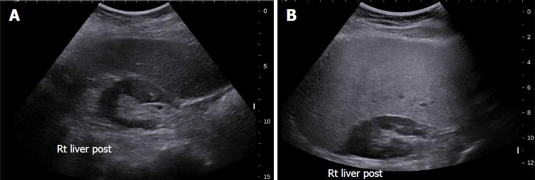
Figure 2 Grey-scale ultrasound in non-alcoholic fatty liver disease.
A: 47-year-old female with increased echogenicity of the liver relative to the right kidney, a classic sonographic finding of hepatic steatosis. The patient had elevated serum liver enzymes and underwent a liver biopsy for NASH evaluation; B: 51-year-old female who underwent liver biopsy as part of clinical follow-up. On evaluation of liver pathology, there was no steatosis. Ultrasound image shows normal echogenicity of the liver parenchyma, which is only slightly hyperechoic relative to the renal parenchyma.
- Citation: Li Q, Dhyani M, Grajo JR, Sirlin C, Samir AE. Current status of imaging in nonalcoholic fatty liver disease. World J Hepatol 2018; 10(8): 530-542
- URL: https://www.wjgnet.com/1948-5182/full/v10/i8/530.htm
- DOI: https://dx.doi.org/10.4254/wjh.v10.i8.530


