Copyright
©The Author(s) 2017.
World J Hepatol. Mar 28, 2017; 9(9): 477-486
Published online Mar 28, 2017. doi: 10.4254/wjh.v9.i9.477
Published online Mar 28, 2017. doi: 10.4254/wjh.v9.i9.477
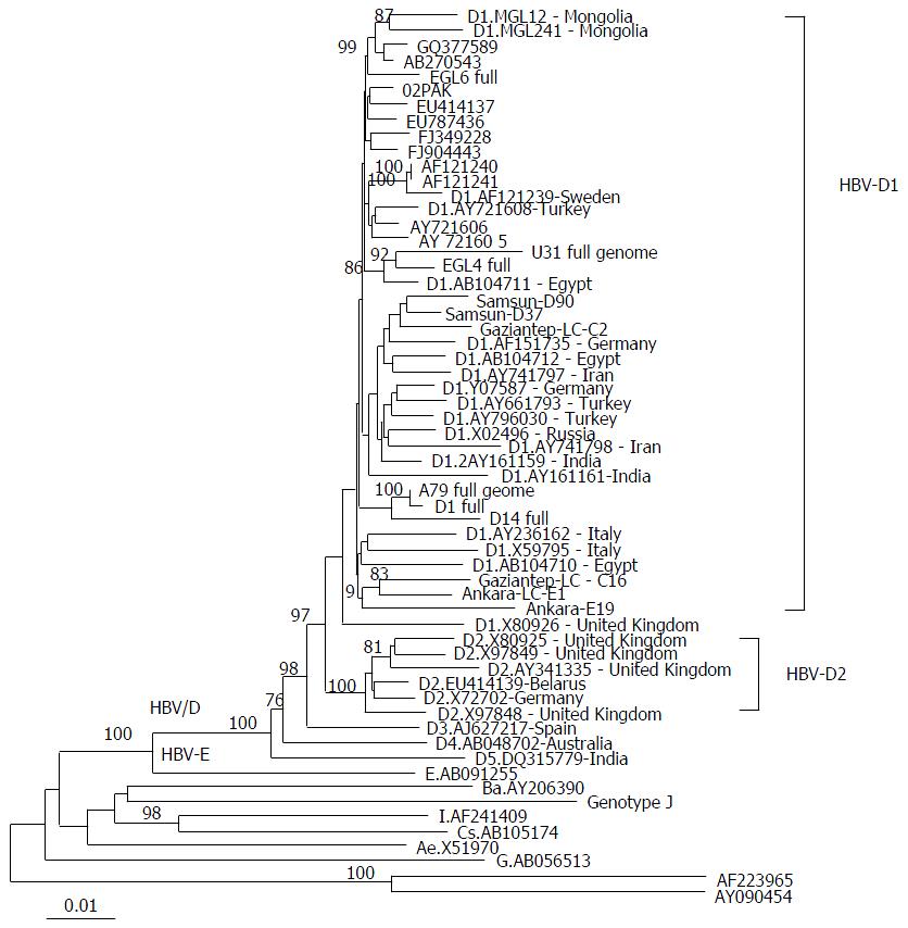
Figure 1 Phylogenetic tree constructed from the nucleotide sequences of the full hepatitis B virus genome.
The phylogenetic tree is constructed by the neighbor joining method and significant bootstrap values (> 75%) are indicated in the tree roots. HBV sequences isolated from the studied cohort are indicated in bold. Reference sequences retrieved from the GenBank/EMBL/DDBJ are indicted by their accession numbers. The country origin of the reference sequences is indicated in brackets. HBV genotypes A-H are indicated in the cluster roots. HBV: Hepatitis B virus.
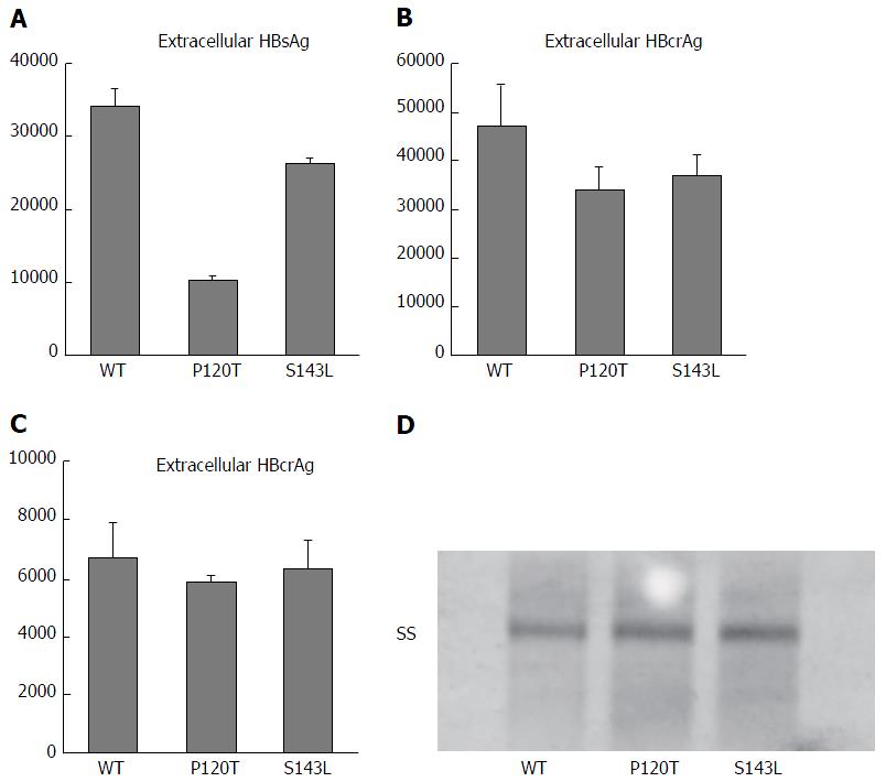
Figure 2 Expression of hepatitis B virus DNA and antigens after transfection of Huh 7 cells with wild type hepatitis B virus genotype D clone (WT) and mutants with S gene mutations P120T (120T) and S143L (143L).
A and B represent the extracellular expression of HBsAg and HBcrAg, respectively, by WT and S gene mutants (120T and 143L); C represents the intracellular expression of the core gene product (HBcrAg) by WT and S gene mutant clones; D: The density of the single-stranded DNA in Southern blot analysis of cell lysates of Huh7 cells transfected with WT and mutants with S gene mutations.
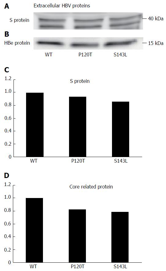
Figure 3 Western blot analysis of hepatitis B surface antigen (A, C), hepatitis B virus core (B), core related proteins (D) expressed by hepatitis B virus genotype D clones (WT) and hepatitis B surface antigen mutant clones (120T and 143L) in the supernatant of Huh 7 cells.
The results for the wild type clones in the three independent transfection experiments were similar and their mean value is set at 1.0. The hepatitis B surface antigen and hepatitis B virus core proteins levels are expressed relative to this value in (C) and (D). HBV: Hepatitis B virus.
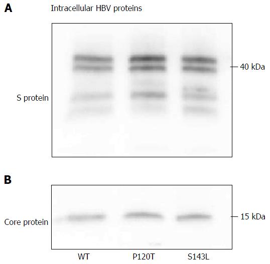
Figure 4 Western blot analysis of hepatitis B surface antigen (A) and hepatitis B virus core (B) proteins expressed by hepatitis B virus genotype D clones (WT) and hepatitis B surface antigen mutant clones (120T and 143L) in the cell lysate of Huh 7 cells.
HBV: hepatitis B virus.
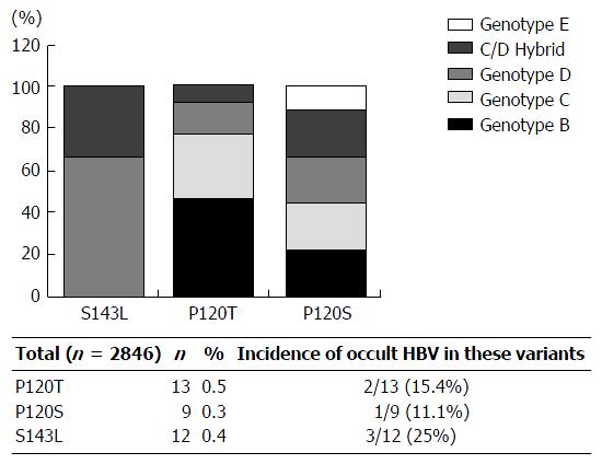
Figure 5 Genotype distribution of the detected S gene product mutation among the database reference sequences.
HBV: Hepatitis B virus.
- Citation: Elkady A, Iijima S, Aboulfotuh S, Mostafa Ali E, Sayed D, Abdel-Aziz NM, Ali AM, Murakami S, Isogawa M, Tanaka Y. Characteristics of escape mutations from occult hepatitis B virus infected patients with hematological malignancies in South Egypt. World J Hepatol 2017; 9(9): 477-486
- URL: https://www.wjgnet.com/1948-5182/full/v9/i9/477.htm
- DOI: https://dx.doi.org/10.4254/wjh.v9.i9.477


