Copyright
©The Author(s) 2017.
World J Hepatol. Feb 28, 2017; 9(6): 343-348
Published online Feb 28, 2017. doi: 10.4254/wjh.v9.i6.343
Published online Feb 28, 2017. doi: 10.4254/wjh.v9.i6.343
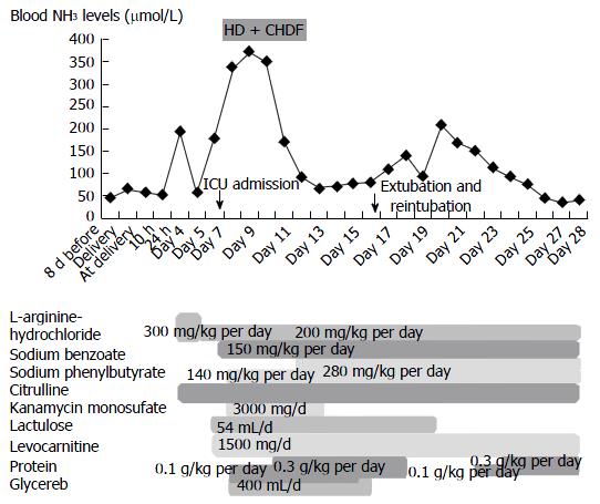
Figure 1 Blood ammonia levels after delivery.
The patient was admitted on emergency basis because of impaired consciousness 7 d after delivery (day 7) and was intubated. She then underwent HD and CHDF under ICU management (from day 8 to day 12). HD: Hemodialysis; CHDF: Continuous hemodiafiltration; ICU: Intensive care unit.
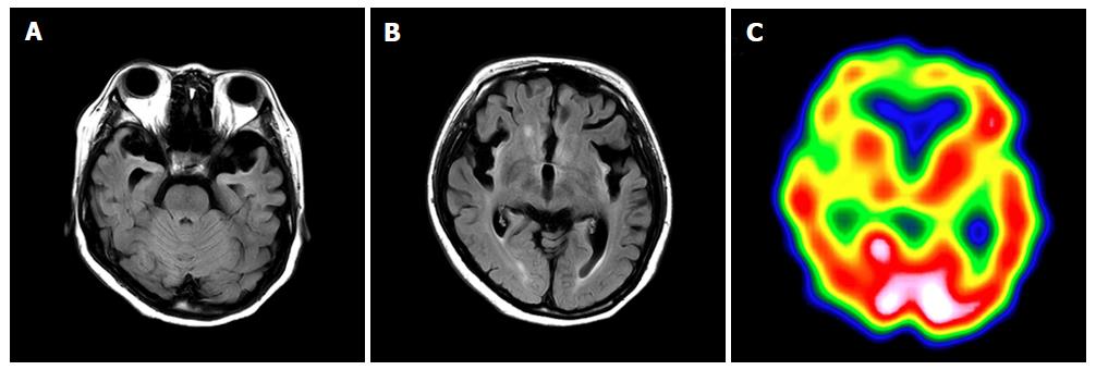
Figure 2 Brain magnetic resonance imaging and single-photon emission computed tomography 7 mo after delivery.
A and B: FLAIR demonstrates atrophy of the bilateral frontal and temporal lobes, and a high signal for the cortex and subcortex on both sides of insula, the ventral tamporal lobe and the frontal lobe bottom. The signal is elevated bilaterally in the putamen, caudate nucleus, and front globus pallidus; C: Single-photon emission computed tomography shows bilaterally decreased blood flow in the frontal lobe, ventral temporal lobe, basal ganglia, and thalamus.
- Citation: Kido J, Kawasaki T, Mitsubuchi H, Kamohara H, Ohba T, Matsumoto S, Endo F, Nakamura K. Hyperammonemia crisis following parturition in a female patient with ornithine transcarbamylase deficiency. World J Hepatol 2017; 9(6): 343-348
- URL: https://www.wjgnet.com/1948-5182/full/v9/i6/343.htm
- DOI: https://dx.doi.org/10.4254/wjh.v9.i6.343


