Copyright
©The Author(s) 2017.
World J Hepatol. Feb 28, 2017; 9(6): 300-309
Published online Feb 28, 2017. doi: 10.4254/wjh.v9.i6.300
Published online Feb 28, 2017. doi: 10.4254/wjh.v9.i6.300
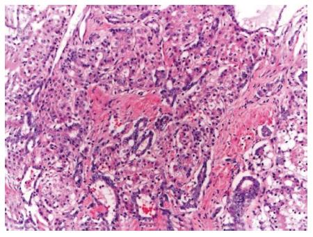
Figure 1 Representative picture classic type combined hepatocellular-cholangiocarcinoma, hematoxylin and eosin, 10 × - intermediate areas with both hepatocytic and chloangiocytic components.
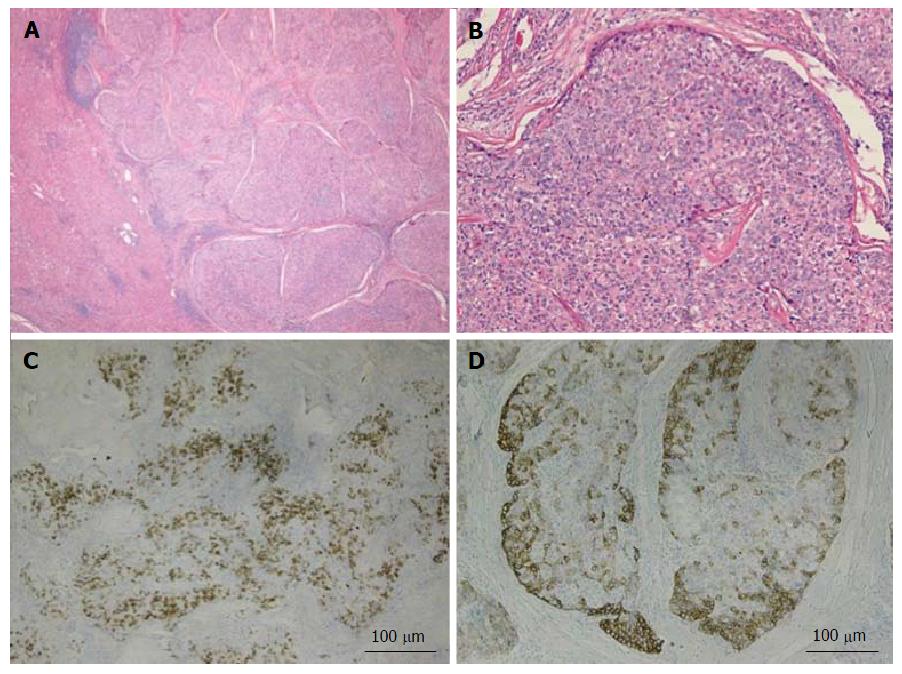
Figure 2 Combined hepatocellular-cholangiocarcinoma with stem cell features, typical subtype.
A: H and E, 4 × - tumor nests present on the right side with non-neoplastic liver on the left side; B: H and E, 10 × - peripheral small cells with hyperchromatic nuclei with mature appearing hepatocytes in the center; C: CK7, 4 × - scattered expression of CK7 by tumor cells; D: CK19, 4 × - patchy staining of the tumor and highlighting small tumor cells located at the periphery. Tumor was also positive for Hep-Par1 (not shown). H and E: Hematoxylin and eosin.
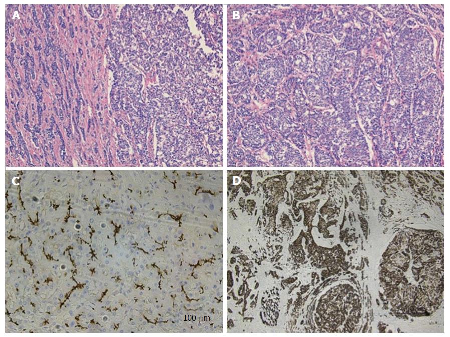
Figure 3 Combined hepatocellular-cholangiocarcinoma with stem cell features, intermediate subtype.
A: H and E, 4 × - tumor is present in trabecular/nested pattern on the right side with ill-formed gland like structures seen on the left side; B: H and E, 10 × - tumor cells with intermediate features between hepatocytes and cholangiocytes; C: CD10, 10 × - tumor showing canalicular staining pattern for CD10 (hepatocytic marker); D: CK19, 4 × - tumor cells strongly and diffusely expressing CK19 (chlolangiocytic marker). Focal tumor cells were positive for Hep-Par1 and CD56 (not shown). H and E: Hematoxylin and eosin.
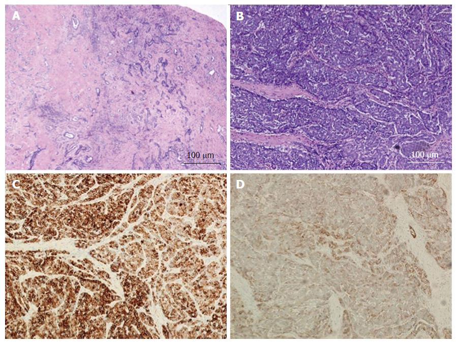
Figure 4 Combined hepatocellular-cholangiocarcinoma with stem cell features, cholangiocellular subtype.
A: H and E, 4 × - tumor cells present in tubular, anastomosing (antler-like) pattern; B: H and E, 10 × - small hyperchromatic tumor cells with high nuclear to cytoplasmic ratio present within dense fibrous stroma; C: CK7, 10 × - tumor is diffusely positive for CK7; D: CD56, 10 × - CD56 staining the cholangiolocellular component as well as the tumor cells at the periphery of the trabeculae. The tumor was diffusely positive for CK19 while negative for HepPar-1 and AFP (not shown). H and E: Hematoxylin and eosin.
- Citation: Gera S, Ettel M, Acosta-Gonzalez G, Xu R. Clinical features, histology, and histogenesis of combined hepatocellular-cholangiocarcinoma. World J Hepatol 2017; 9(6): 300-309
- URL: https://www.wjgnet.com/1948-5182/full/v9/i6/300.htm
- DOI: https://dx.doi.org/10.4254/wjh.v9.i6.300


