Copyright
©The Author(s) 2017.
World J Hepatol. Sep 18, 2017; 9(26): 1081-1091
Published online Sep 18, 2017. doi: 10.4254/wjh.v9.i26.1081
Published online Sep 18, 2017. doi: 10.4254/wjh.v9.i26.1081
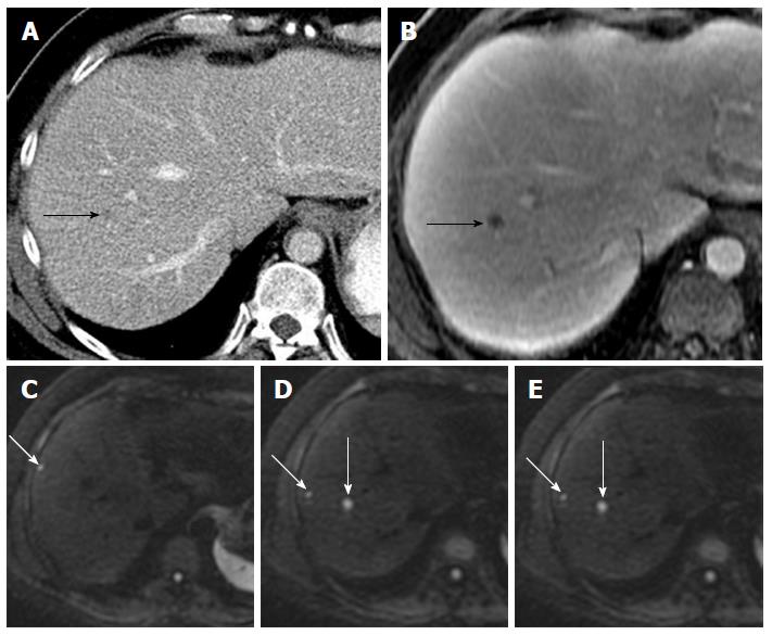
Figure 1 Value of diffusion-weighted magnetic resonance imaging in lesion detection in a 51-year-old male with metastatic leiomyosarcoma of the thigh.
A: Axial contrast enhanced CT scan demonstrated a subtle hypodensity in the right lobe of liver (black arrow); B: Axial post gadolinium T1-weighted MR image demonstrates a single metastatic lesion (black arrow); C-E: DW-MR image at b-600 demonstrates additional lesions (white arrows). DW-MR: Diffusion-weighted magnetic resonance; CT: Computed tomography.
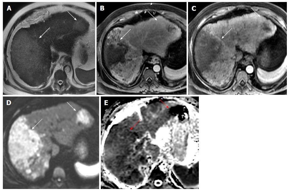
Figure 2 A 66-year-old lady with multifocal infiltrative hepatocellular carcinoma with improved detection on diffusion-weighted imaging.
(A) Axial T2 weighted image demonstrates multifocal areas of T2 hyperintense masses (white arrows) which demonstrate heterogeneous arterial hyperenhancement on post gadolinium late arterial phase images (B) and washout appearance on portal venous phase images (C). (D) Axial DWI image at b-600 and (E) ADC image show that these masses demonstrate restricted diffusion and are better appreciated than the dynamic phase images. Serum Alpha feto-protein value of 1552. DWI: Diffusion-weighted imaging.
- Citation: Shenoy-Bhangle A, Baliyan V, Kordbacheh H, Guimaraes AR, Kambadakone A. Diffusion weighted magnetic resonance imaging of liver: Principles, clinical applications and recent updates. World J Hepatol 2017; 9(26): 1081-1091
- URL: https://www.wjgnet.com/1948-5182/full/v9/i26/1081.htm
- DOI: https://dx.doi.org/10.4254/wjh.v9.i26.1081


