Copyright
©The Author(s) 2017.
World J Hepatol. Aug 28, 2017; 9(24): 1022-1029
Published online Aug 28, 2017. doi: 10.4254/wjh.v9.i24.1022
Published online Aug 28, 2017. doi: 10.4254/wjh.v9.i24.1022
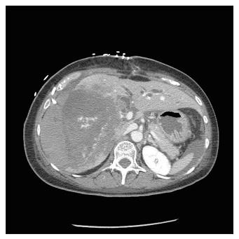
Figure 1 Computed tomography scan show a massive intrahepatic hematoma involving the right hepatic lobe and segment IV.
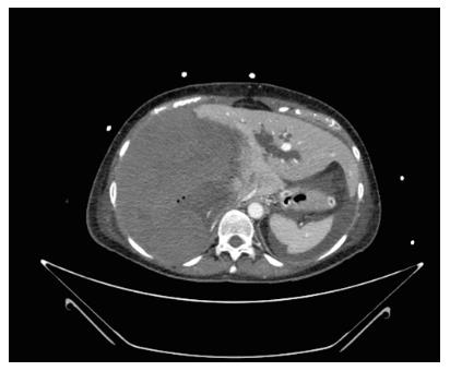
Figure 2 Computed tomography scan show the ischemic liver necrosis after the right hepatic artery embolization.
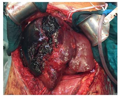
Figure 3 Intraoperative findings: A huge right lobe hematoma extended to segment IV with signs of extrahepatic rupture.
- Citation: Lauterio A, De Carlis R, Di Sandro S, Ferla F, Buscemi V, De Carlis L. Liver transplantation in the treatment of severe iatrogenic liver injuries. World J Hepatol 2017; 9(24): 1022-1029
- URL: https://www.wjgnet.com/1948-5182/full/v9/i24/1022.htm
- DOI: https://dx.doi.org/10.4254/wjh.v9.i24.1022


