Copyright
©The Author(s) 2017.
World J Hepatol. Jul 8, 2017; 9(19): 840-849
Published online Jul 8, 2017. doi: 10.4254/wjh.v9.i19.840
Published online Jul 8, 2017. doi: 10.4254/wjh.v9.i19.840
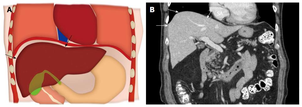
Figure 1 Anatomy of the hepatic dome.
A: Colored schematic diagram; B: Coronal reformatted computed tomography image demonstrating the anatomy of the hepatic dome (arrows).
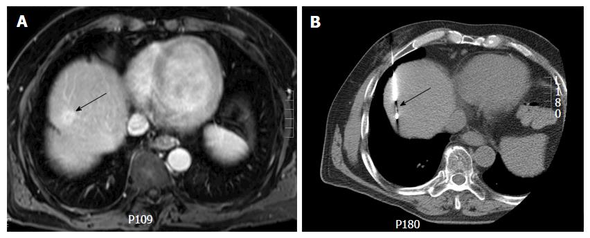
Figure 2 Computed tomography guided biopsy of a liver dome lesion in a 61-year-old man.
A: Axial post gadolinium T1-weighted magnetic resonance image shows a 2 cm lesion (arrow) in the hepatic dome. On pre-procedural computed tomography, the tumor was not well seen and contrast could not be administered due to iodine allergy; B: Needle placement for biopsy was done based on use of anatomic landmarks (arrow) (configuration of inferior vena cava, cardiac margin and aorta) via a transpulmonary approach. Histopathology: Hepatocellular carcinoma.
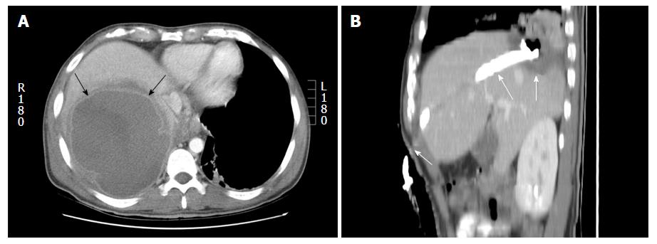
Figure 3 Percutaneous drainage of hepatic dome abscess in a 36-year-old man.
A: Axial contrast computed tomography shows the large hepatic dome abscess (arrows). Pleural transgression carried an increased risk of pleural complications; B: Percutaneous catheter drainage using a subcostal approach (arrows) allowed successful abscess treatment while avoiding pleural transgression.
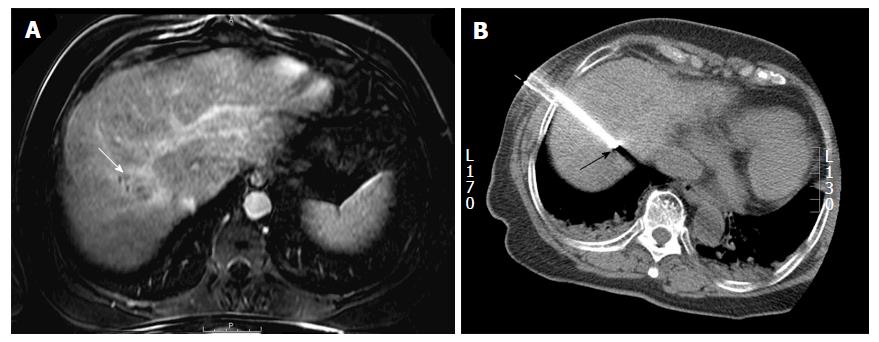
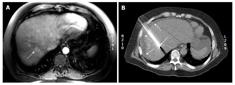
Figure 4 Percutaneous radiofrequency ablation of a hepatic dome hepatocellular carcinoma in a 54-year-old man.
A: Axial post gadolinium T1-weighted image in the portal venous phase demonstrates a 3.4 cm hepatocellular carcinoma in the hepatic dome (arrow); B: During the radiofrequency ablation procedure, the patient was placed in the oblique position and using a lateral intercostal approach the tumor was accessed for a successful ablation (arrow).
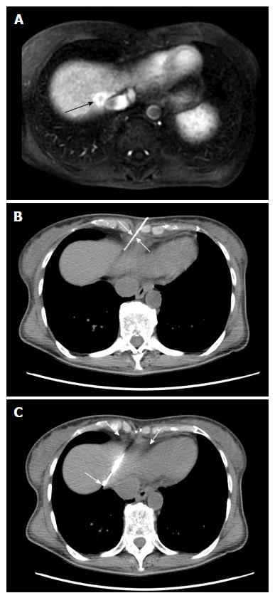
Figure 5 Computed tomography guided biopsy of a liver lesion adjacent to the inferior vena cava in a 56-year-old woman with breast cancer.
A: Axial post gadolinium image T1 weighted magnetic resonance image demonstrates an enhancing lesion adjacent to the inferior vena cava (arrow); B and C: Intraprocedural computed tomography images demonstrate placement of the biopsy needle into the lesion through the epipericardial fat pad (arrows). Biopsy: Breast cancer metastases.
Figure 6 Computed tomography guided radiofrequency ablation in a 56-year-old lady with colorectal liver metastases.
A: Axial post gadolinium T1 weighted magnetic resonance image shows a 2.7 cm (arrow) hepatic dome metastases; B: The radiofrequency ablation was performed with the patient in supine position and needle placement through the anterolateral intercostal approach. Hydrodissection was performed in this patient (arrows).
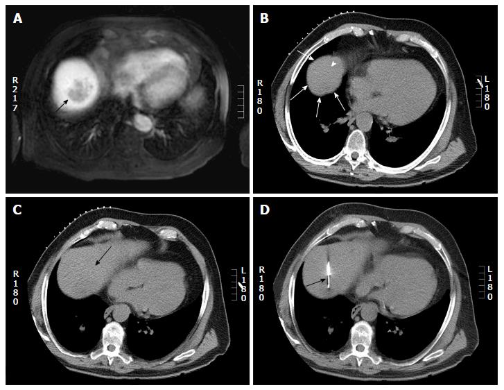
Figure 7 Computed tomography guided biopsy in a 65-year-old man.
A: Axial post gadolinium magnetic resonance imaging shows a 4 cm hepatic dome lesion; B: On preprocedural CT, the lesion in the high dome is surrounded by lung (arrow head) on all sides. Pulmonic transgression was not possible as the patient had severe emphysema; C: The CT gantry was angulated in the craniocaudal direction (20 degrees) which created a safe path to the tumor from the anterior aspect (arrow); D: Axial intraprocedural CT image shows biopsy needle within the lesion (arrow). Biopsy: Hepatocellular carcinoma. CT: Computed tomography.
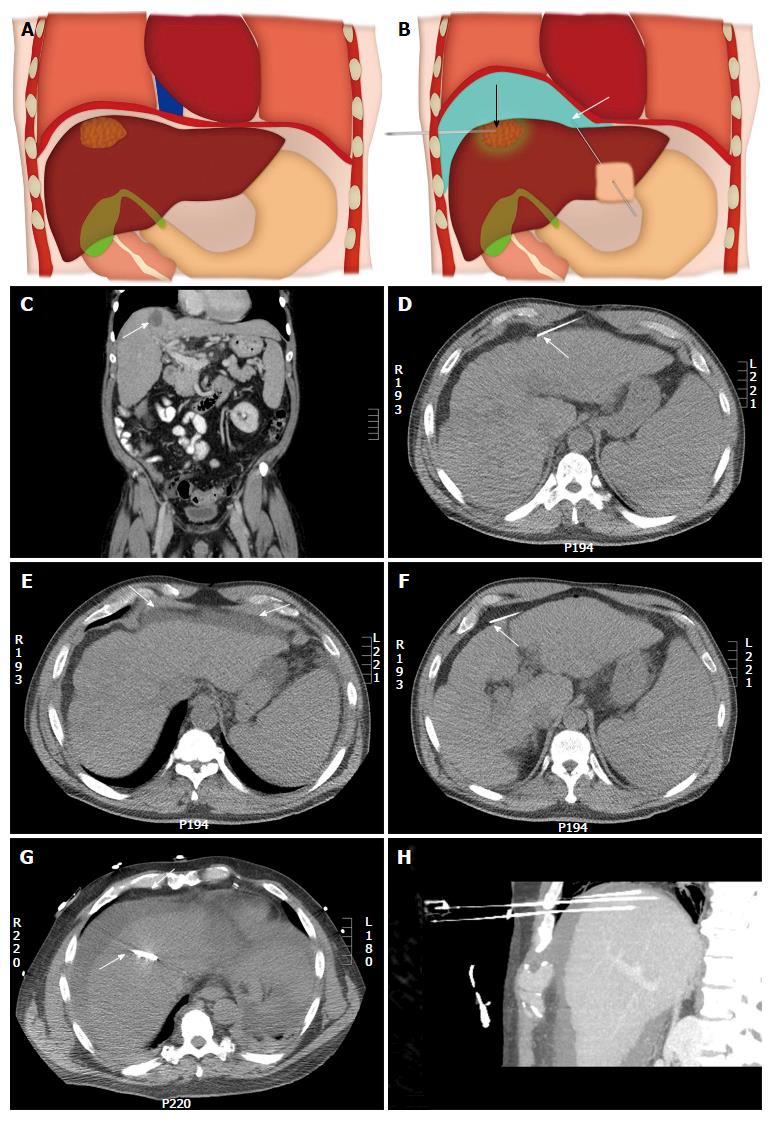
Figure 8 Illustration of the upper abdomen demonstrating the hydro-dissection technique.
A: Coronal colored image shows a hepatic dome lesion very close to the diaphragm; B: Coronal colored image after hydro-dissection (shown in blue color) shows the separation of the dome of liver from the diaphragm which improves percutaneous access to the lesion and limits diaphragmatic injury. White arrow shows the needle for hydodissection and black arrow shows the needle into the lesion. An example of a hepatic dome lesion (C) (arrow) where hydrodissection was attempted by needle placed anteriorly (D) (arrow); E: Axial computed tomography showed accumulation of fluid within the properitoneal fat (arrows); F: The needle was repositioned with the tip of the needle into the peritoneal cavity (arrow); G: Successful hydrodissection achieved using instillation of 500 cc of D5W through the needle (arrow); H: The fluid was used to create a safe path for radiofrequency ablation of the hepatic dome hepatocellular carcinoma. Sagittal Maximum intensity projection image demonstrating the artificial ascites and electrodes in position in the hepatic dome lesion.
Figure 9 Artificial pneumothorax for radiofrequency ablation of hepatic dome hepatocellular carcinoma in a 69-year-old man.
A: Axial T2WI magnetic resonance imaging shows A 4 cm lesion (arrow) in the hepatic dome; B: Artificial pneumothorax was created after instillation of intrapleural air. A chest tube was placed for drainage (arrow); C: Intra-procedural computed tomography shows radiofrequency electrode within the lesion for a successful ablation (black arrow).
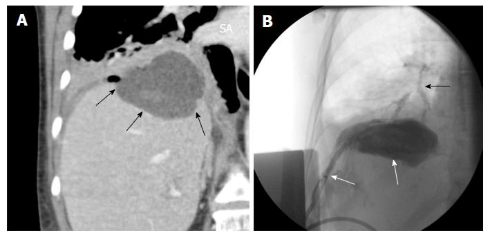
Figure 10 Hepatic abscess complicating a hepatic dome metastases ablation.
A: Coronal reformatted image shows a abscess in the dome of liver (arrows); B: Percutaneous drainage was performed and drain injection shows communication with the bronchi (black arrow).
- Citation: Kambadakone A, Baliyan V, Kordbacheh H, Uppot RN, Thabet A, Gervais DA, Arellano RS. Imaging guided percutaneous interventions in hepatic dome lesions: Tips and tricks. World J Hepatol 2017; 9(19): 840-849
- URL: https://www.wjgnet.com/1948-5182/full/v9/i19/840.htm
- DOI: https://dx.doi.org/10.4254/wjh.v9.i19.840


