Copyright
©The Author(s) 2016.
World J Hepatol. Dec 8, 2016; 8(34): 1535-1540
Published online Dec 8, 2016. doi: 10.4254/wjh.v8.i34.1535
Published online Dec 8, 2016. doi: 10.4254/wjh.v8.i34.1535
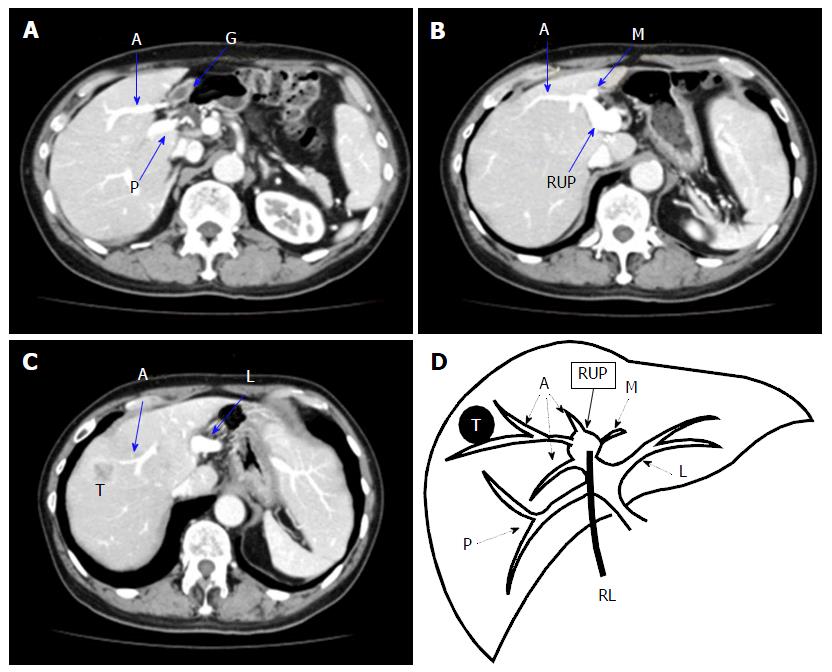
Figure 1 Case 1 enhanced computed tomography.
A: Computed tomography shows the left-sided gallbladder and RUP; B: The right anterior and medial segmental portal branches ramify from the RUP after its trifurcation as well as the right posterior and left lateral branch; C: A 25-mm sized tumour peripherally enhanced in the arterial phase was detected in segment 8; D: Diagram of the intrahepatic portal vein branching and the location of the tumour. A: Right anterior portal vein; P: Right posterior portal vein; G: Gallbladder; M: Left medial portal vein; RUP: Right umbilical portion; L: Left lateral portal vein; T: Tumour; RL: Round ligament.
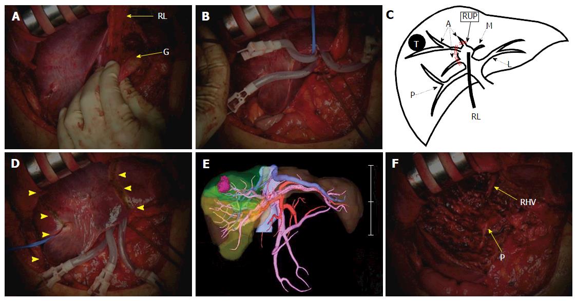
Figure 2 Case 1 operative findings.
A: The gallbladder was attached to the round ligament; B: Three ramifications of the right anterior Glissonean pedicles were separated and clamped; C: Diagram of the clamped Glissonean pedicles (double line); D and E: The demarcation area (arrow head) was identified as in the preoperative simulation; F: The accomplishment of a right anterior sectionectomy. RL: Round ligament; G: Gallbladder; A: Right anterior branch of the Glissonean pedicle; P: Right posterior branch of the Glissonean pedicle; M: Left medial branch of the Glissonean pedicle; RUP: Right umbilical portion; L: Left lateral branch of the Glissonean pedicle; T: Tumour; RHV: Right hepatic vein.
Figure 3 Case 2 enhanced computed tomography.
A and B: CT shows the right posterior portal branch to be solely bifurcated, and the right anterior and medial segmental portal branches ramify from the RUP; B: A 25-mm sized mass (arrow head) is adjacent to the RUP. The RUP is almost occluded, and the intrahepatic distal bile duct is dilated (B); C: Diagram of the intrahepatic portal vein branching and the location of the tumour. RL: Round ligament; G: Gallbladder; A: Right anterior branch of the Glissonean pedicle; P: Right posterior branch of the Glissonean pedicle; M: Left medial branch of the Glissonean pedicle; RUP: Right umbilical portion; L: Left lateral branch of the Glissonean pedicle; T: Tumour; RHV: Right hepatic vein.
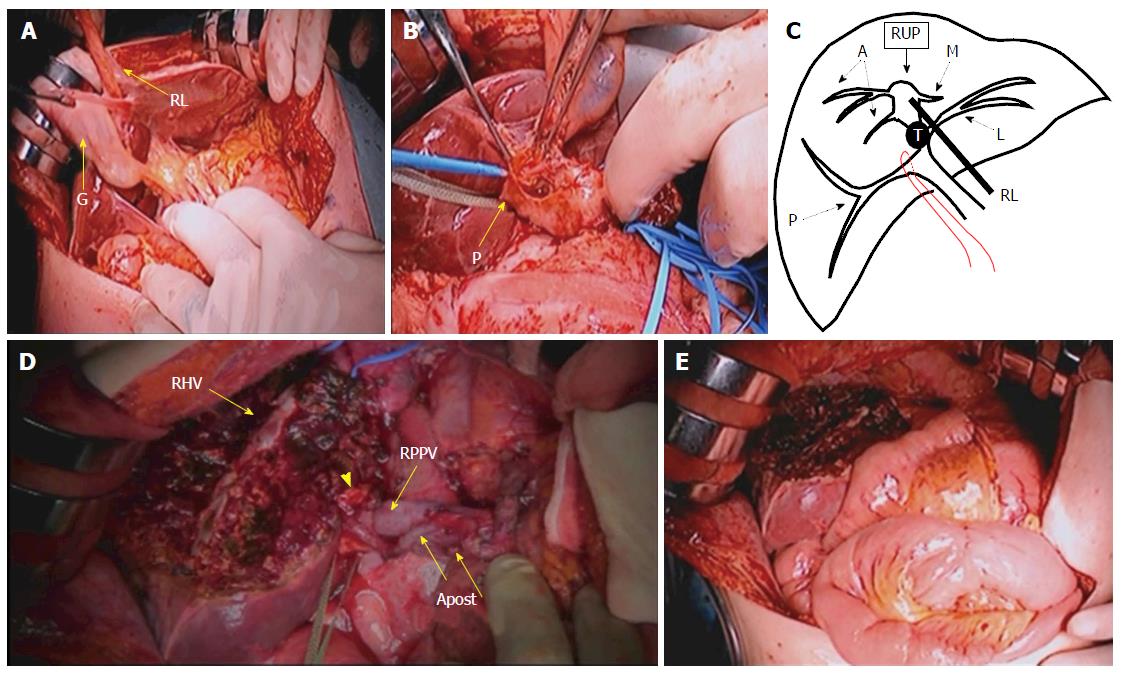
Figure 4 Case 2 operative findings.
A: The gallbladder was attached to the round ligament; B: The right posterior Glissonean pedicle was encircled, and the vessels entering the right posterior Glissonean pedicle were identified; C: Diagram of securing the right posterior branch of the Glissonean pedicle; D: The accomplishment of left trisectionectomy; E: Hepaticojejunostomy was performed. RL: Round ligament; G: Gallbladder; A: Right anterior branch of the Glissonean pedicle; P: Right posterior branch of the Glissonean pedicle; M: Left medial branch of the Glissonean pedicle; RUP: Right umbilical portion; L: Left lateral branch of the Glissonean pedicle; T: Tumour; RHV: Right hepatic vein; RPPV: Right posterior portal vein; Apost: Right posterior hepatic artery; Arrow-head: Stump of the right posterior bile duct.
- Citation: Ome Y, Kawamoto K, Park TB, Ito T. Major hepatectomy using the glissonean approach in cases of right umbilical portion. World J Hepatol 2016; 8(34): 1535-1540
- URL: https://www.wjgnet.com/1948-5182/full/v8/i34/1535.htm
- DOI: https://dx.doi.org/10.4254/wjh.v8.i34.1535


