Copyright
©The Author(s) 2016.
World J Hepatol. Jun 28, 2016; 8(18): 779-784
Published online Jun 28, 2016. doi: 10.4254/wjh.v8.i18.779
Published online Jun 28, 2016. doi: 10.4254/wjh.v8.i18.779
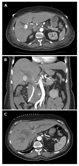
Figure 1 Computerized tomography scan demonstrating pseudoaneurysm and significant biliary ductal dilatation.
A: Triple contrast computerized tomography scan of abdomen demonstrates hepatic artery pseuodoaneurysm; B: With significant thrombus formation adjacent to aneurysm; C: Significant biliary ductal dilatation in the right and left hepatic ducts.
Figure 2 Interventional attempts to embolize the hepatic artery pseudoaneurysm.
A: Angiogram of common hepatic artery demonstrates pseudoaneurysmal formation off right hepatic artery; B and C: Interventional fluoroscopic and ultrasound-guided embolization of right hepatic artery pseudoaneurysm with multiple coils, gelfoam, and thrombin.
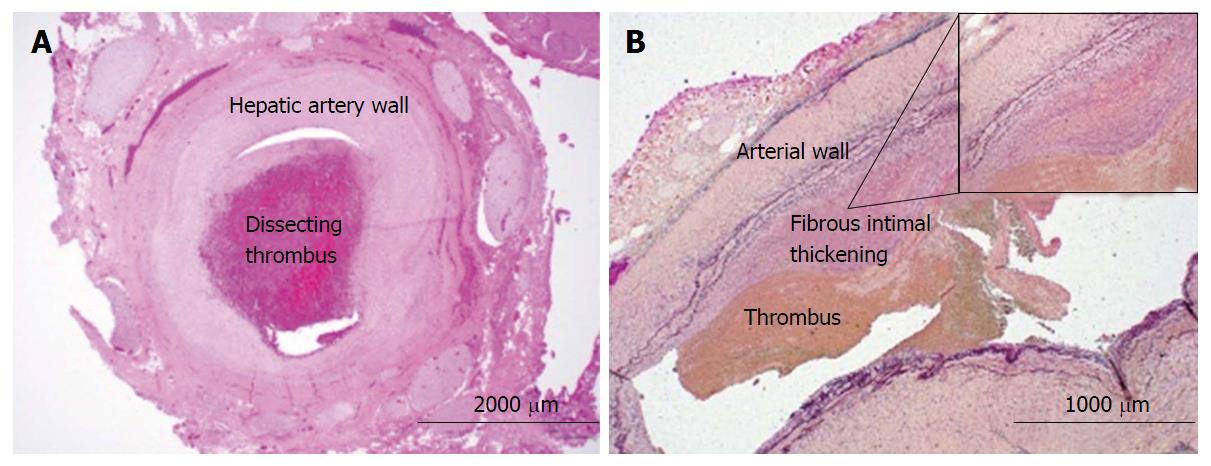
Figure 3 Histochemical analyses of resected hepatic artery.
A: H and E of hepatic artery with organized thrombus dissecting into the vessel wall, 40 × magnification; B: Verhoeff-Van Gieson (VVG) staining of the hepatic artery with eccentric fibrous intimal thickening with adherent thrombus, 20 × magnification (B, inlet). Internal elastic lamina is highlighted by the VVG stain with notable fibrous intimal thickening, 100 × magnifications.
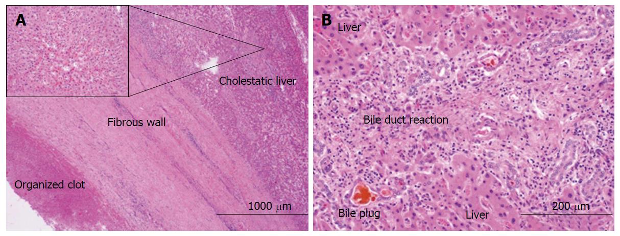
Figure 4 Histologic analyses of hepatic artery pseudoaneurysm and adjacent liver.
A: H and E of fibrous capsule of the contained pseudoaneurysmal rupture adjacent to liver, 20 × magnification (A, inlet). Liver with centrilobular cholestasis, 200 × magnification; B: Liver with bile duct reaction and bile plugs consistent with biliary obstruction, 200 × magnification.
- Citation: Luckhurst CM, Perez C, Collinsworth AL, Trevino JG. Atypical presentation of a hepatic artery pseudoaneurysm: A case report and review of the literature. World J Hepatol 2016; 8(18): 779-784
- URL: https://www.wjgnet.com/1948-5182/full/v8/i18/779.htm
- DOI: https://dx.doi.org/10.4254/wjh.v8.i18.779


