Copyright
©The Author(s) 2015.
World J Hepatol. Feb 27, 2015; 7(2): 127-138
Published online Feb 27, 2015. doi: 10.4254/wjh.v7.i2.127
Published online Feb 27, 2015. doi: 10.4254/wjh.v7.i2.127
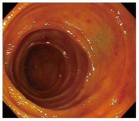
Figure 1 Mucosal red spots.
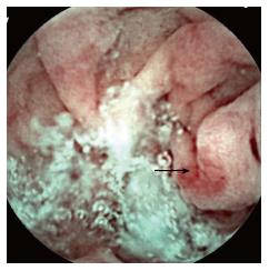
Figure 2 Angiodysplasia-like lesion.
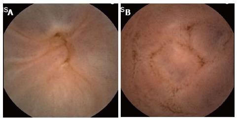
Figure 3 Ileal varices (A and B).
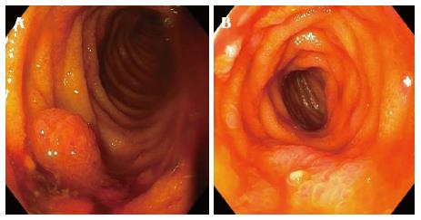
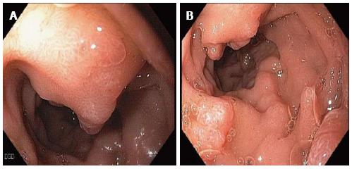
Figure 4 Portal hypertensive polypoid enteropathy (A and B).
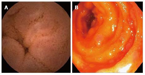
Figure 5 Herring roe appearance of small bowel mucosa (A and B).
Figure 6 Mucosal edema with granularity of the small bowel (A and B).
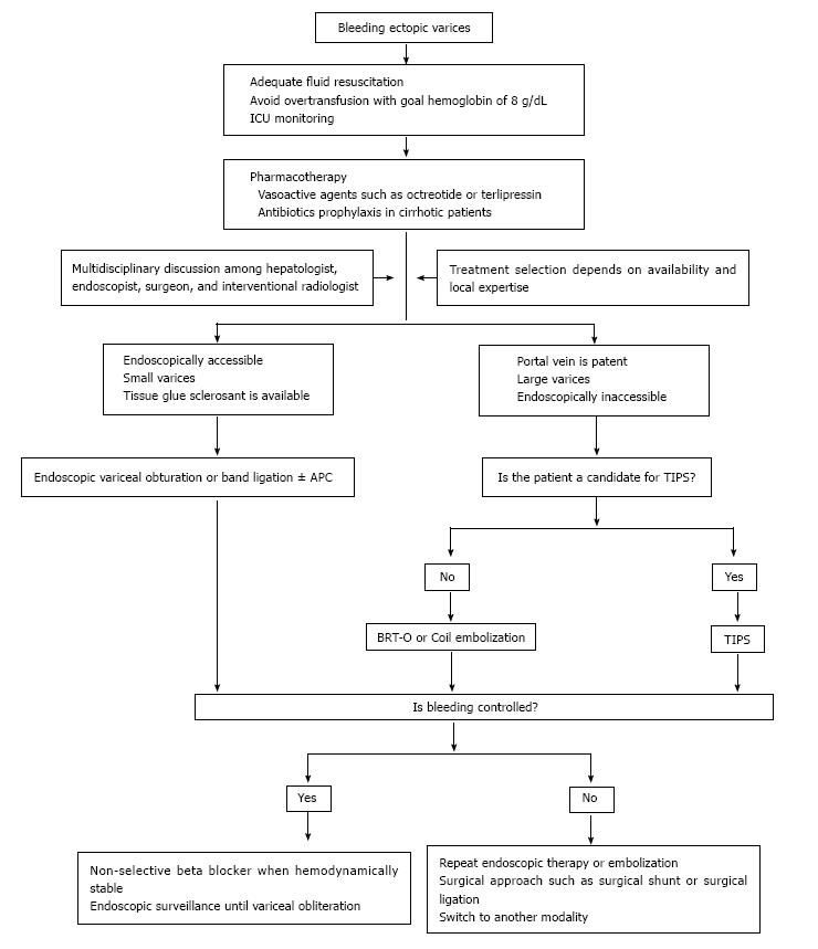
Figure 7 Management of bleeding ectopic varices.
TIPS: Transjugular intrahepatic portosystemic shunt; APC: Argon plasma coagulation; BRTO: Balloon-occluded retrograde transvenous obliteration.
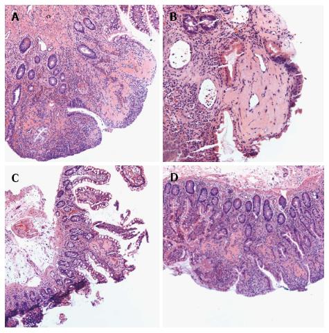
Figure 8 Histopathological changes of portal hypertensive enteropathy (A-D).
- Citation: Mekaroonkamol P, Cohen R, Chawla S. Portal hypertensive enteropathy. World J Hepatol 2015; 7(2): 127-138
- URL: https://www.wjgnet.com/1948-5182/full/v7/i2/127.htm
- DOI: https://dx.doi.org/10.4254/wjh.v7.i2.127


