Copyright
©The Author(s) 2015.
World J Hepatol. Jul 18, 2015; 7(14): 1856-1865
Published online Jul 18, 2015. doi: 10.4254/wjh.v7.i14.1856
Published online Jul 18, 2015. doi: 10.4254/wjh.v7.i14.1856
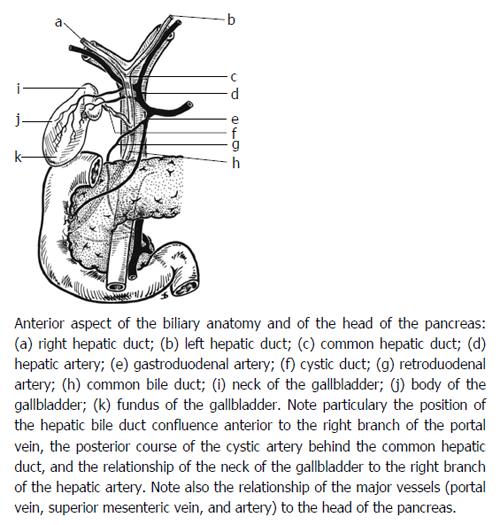
Figure 1 Arterial supply of the biliary tree[19] (reprinted with permission from Elsevier).
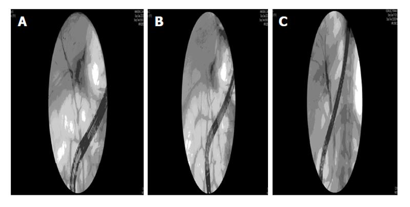
Figure 2 Endoscopic retrograde cholangiograms from a patient with an anastomotic leak after live donor liver transplantation.
A: Cholangiogram demonstrating leak (extravasation of contrast) coming off the anastomosis after right lobe live donor liver transplant; B: Cholangiogram with plastic stent deployed across the anastomosis to heal the leak; C: Cholangiogram showing resolution of the leak several months later.
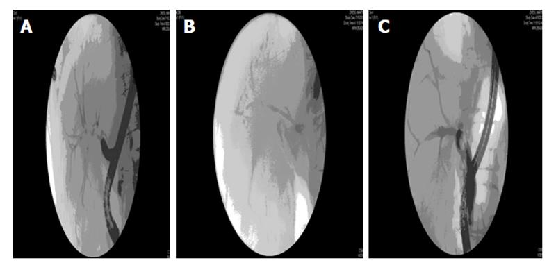
Figure 3 Endoscopic retrograde cholangiograms from a patient with an anastomotic stricture after live donor liver transplantation.
A: Cholangiogram demonstrating complex anastomotic stricture after right lobe live donor liver transplant; B: Cholangiogram with plastic stent deployed across the stricture; C: Cholangiogram showing marked improvement in stricture after multiple dilation and stenting.
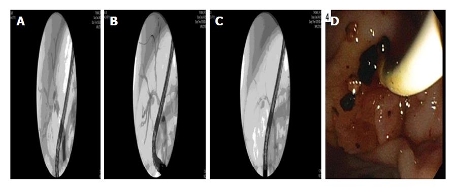
Figure 4 Endoscopic retrograde cholangiograms from a patient with a non-anastomotic stricture after live donor liver transplantation complicated by biliary cast formation (endoscopic image).
A: Cholangiogram demonstrating non-anastomotic stricture after right lobe live donor liver transplant with irregular filling defects (casts) in a dilated segment (running at 8 o’clock in the image); B: Cholangiogram demonstrating clearance of the filling defects; C: Cholangiogram demonstrating two plastic stents deployed into the right anterior and right posterior systems after the casts were removed. Note how well the biliary tree has drained; D: Endoscopic image of the cast material being removed through the ampulla.
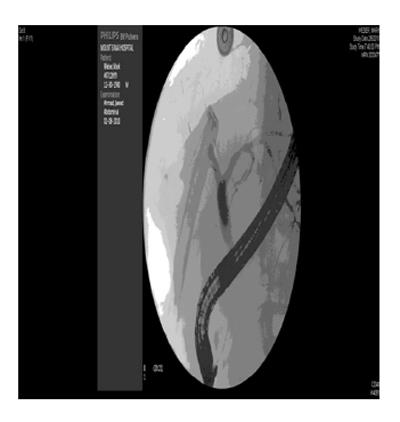
Figure 5 Endoscopic retrograde cholangiogram from a patient with a leak from the remnant right common hepatic duct a few days after right lobe live donor liver transplantation.
The drain to the left can be seen filling when contrast is injected into the right common hepatic duct. This was managed successfully by a transpapillary stent.
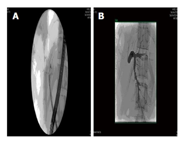
Figure 6 Stricture in donor after right lobe live donor liver transplantation.
A: Endoscopic retrograde cholangiogram showing minimal filling of the left system a few weeks after right lobe live donor liver transplantation; B: Percutaneous transhepatic cholangiogram from the same patient in Figure 4A demonstrating a tight stricture at the take off the left common hepatic duct.
- Citation: Simoes P, Kesar V, Ahmad J. Spectrum of biliary complications following live donor liver transplantation. World J Hepatol 2015; 7(14): 1856-1865
- URL: https://www.wjgnet.com/1948-5182/full/v7/i14/1856.htm
- DOI: https://dx.doi.org/10.4254/wjh.v7.i14.1856


