Copyright
©2014 Baishideng Publishing Group Inc.
World J Hepatol. Aug 27, 2014; 6(8): 559-569
Published online Aug 27, 2014. doi: 10.4254/wjh.v6.i8.559
Published online Aug 27, 2014. doi: 10.4254/wjh.v6.i8.559
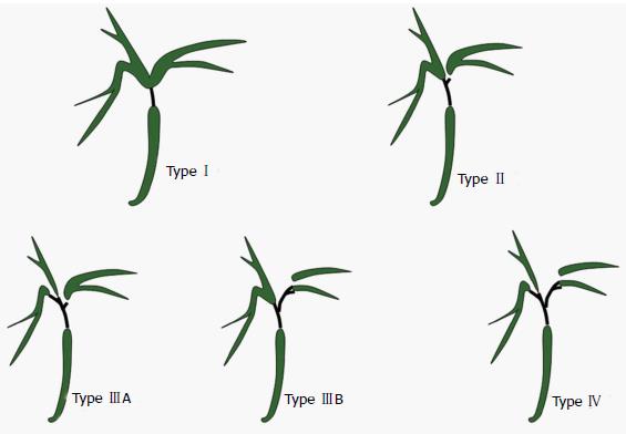
Figure 1 Hilar Cholangicarcinoma-Bismuth classification.
Type I: Confluence of right and left hepatic duct is intact; Type II: Right and left hepatic ducts are separated; Type III: Tumor extends into second degree branches of right (IIIA) or left (IIIB) hepatic duct; Type IV: Involvement of both side secondary branches.
Figure 2 Stenting for hilar cholangiocarcinoma.
A: Unilateral plastic stent; B: Bilateral plastic stents; C: Unilateral metal stent; D: Bilateral metal stents (previously placed percutaneous drains are still in place).
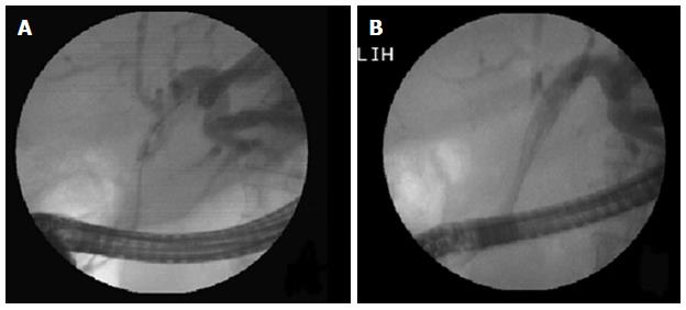
Figure 3 Hilar stricture: Unilateral metal stent placed in dilated left lobe duct.
A: Left duct selectively cannulated and showing dilated system; B: Metal stent being deployed in the left ductal system.
Figure 4 Two metal stents placed side by side in a Type II Bismuth hilar cholangiocarcinoma.
A: Magnetic resonance cholangiopancreaticography showing the stricture; B: Endoscopic view showing two metal stents placed side by side; C: X-ray showing two parallel stents with proximal ends in the right or left ductal systems.
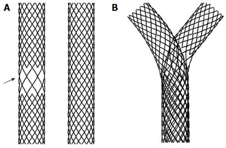
Figure 5 Y stent (stent-in-stent) from Tae Woong Medical (South Korea) for Hilar Cholangiocarcinoma.
A: Two stents one with open mesh in the middle (arrow) and other with normal mesh; B: Y stent system in which the normal mesh stent passes through the open mesh of the second stent.
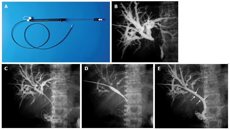
Figure 6 Percutaneous stenting in Bismuth Type I hilar cholangiocarcinoma.
A: Stent assembly; B: Cholangiogram showing the block; C: Guide wire being negotiated across the stricture; D: Balloon dilatation being performed; E: Stent (arrow) after deployment.
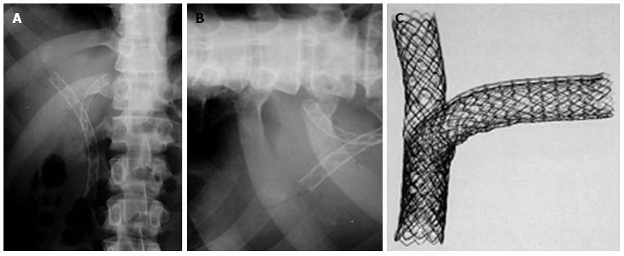
Figure 7 Percutaneous double stenting.
A: X-ray showing the Y configuration of the stent (Boston Scientific MA, United States); B: Transverse limb of a T stent (Taewoong Medial, South Korea) showing the open mesh in the center; C: Assembly of a T stent showing the vertical stent passing through the open mesh of the transverse stent.
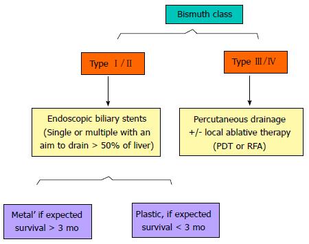
Figure 8 A simplified algorithm suggested for the palliation of Hilar Cholangiocarcinoma, based on Bismuth type.
Additional percutaneous procedures may be required if the endoscopic stenting results are suboptimal. PDT: Photodynamic therapy; RFA: Radiofrequency ablation.
- Citation: Goenka MK, Goenka U. Palliation: Hilar cholangiocarcinoma. World J Hepatol 2014; 6(8): 559-569
- URL: https://www.wjgnet.com/1948-5182/full/v6/i8/559.htm
- DOI: https://dx.doi.org/10.4254/wjh.v6.i8.559


