Copyright
©2014 Baishideng Publishing Group Inc.
World J Hepatol. Oct 27, 2014; 6(10): 745-751
Published online Oct 27, 2014. doi: 10.4254/wjh.v6.i10.745
Published online Oct 27, 2014. doi: 10.4254/wjh.v6.i10.745
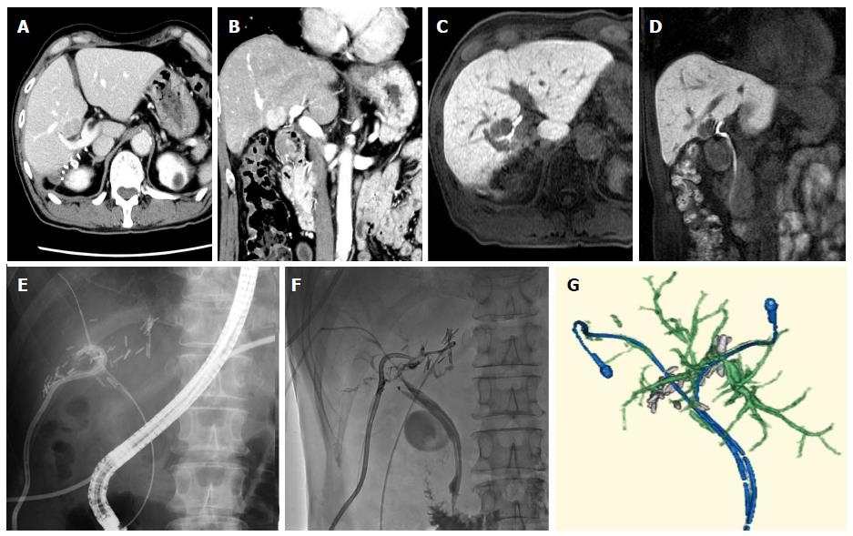
Figure 1 Representative grade C case in bile leakage.
A 67-year-old man had hepatocellular carcinoma (diameter: 2 cm; A: Axial view; B: Coronal view) in segment S5 of his liver (located at the bifurcation of the bile duct in the hilar plate) (C: Axial view; D: Coronal view). The tumor was resected via enucleation; E: Bile leakage was detected and so endoscopic retrograde cholangiodrainage was performed together with percutaneous drainage of the resected pouch; F: Subsequently, stenosis of the left hepatic duct due to bile duct ischemia occurred. Percutaneous transhepatic cholangiodrainage was performed via the B3 duct; G: Three-dimensional reconstruction based on CT images obtained before the patient was discharged from hospital. CT: Computed tomography.
- Citation: Ishii M, Mizuguchi T, Harada K, Ota S, Meguro M, Ueki T, Nishidate T, Okita K, Hirata K. Comprehensive review of post-liver resection surgical complications and a new universal classification and grading system. World J Hepatol 2014; 6(10): 745-751
- URL: https://www.wjgnet.com/1948-5182/full/v6/i10/745.htm
- DOI: https://dx.doi.org/10.4254/wjh.v6.i10.745


