Copyright
©2014 Baishideng Publishing Group Inc.
World J Hepatol. Oct 27, 2014; 6(10): 704-715
Published online Oct 27, 2014. doi: 10.4254/wjh.v6.i10.704
Published online Oct 27, 2014. doi: 10.4254/wjh.v6.i10.704
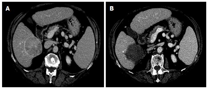
Figure 1 Representative case of complete ablation of large nodule with multi-fibre technique.
A: Computed tomography (CT) scan before laser ablation (LA) session shows a nodular lesion 6 cm in maximum diameter (hepatocellular carcinoma moderately differentiated) localized in the S6 with exophytic growth (exophytic component > 40%); B: CT scan performed 4 wk after LA procedure shows complete necrosis of the tumor. Four illuminations were performed using the pullback technique and the treatment lasted 24 min. The procedure was well tolerated and the patient was discharged from the hospital 24 h after the procedure. The only side effects were mild pain and self-limiting fever lasting for 7 d.
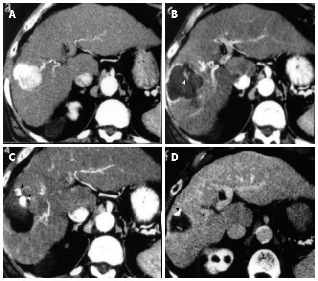
Figure 2 Representative case of complete ablation of hepatocellular carcinoma of 5 cm with combined treatment (laser ablation followed by trans-arterial-chemo-embolization).
A: Computed tomography (CT) scan before Laser ablation (LA) shows a lesion 5 cm in diameter beneath the capsule in S8 during arterial phase; B: CT scan after LA shows an area of necrosis larger than basal lesion with small viable foci (white arrowhead) within the zone of coagulation; C: CT scan shows compact retention of iodized oil in the residual viable tissue (black arrowheads) after trans-arterial-chemo-embolization (TACE) session; D: CT scan shows marked volume reduction of treated area and clear shrinkage of viable tissue (black arrowhead) 6 mo after the combined procedure.
- Citation: Costanzo GGD, Francica G, Pacella CM. Laser ablation for small hepatocellular carcinoma: State of the art and future perspectives. World J Hepatol 2014; 6(10): 704-715
- URL: https://www.wjgnet.com/1948-5182/full/v6/i10/704.htm
- DOI: https://dx.doi.org/10.4254/wjh.v6.i10.704


