Copyright
©2013 Baishideng Publishing Group Co.
World J Hepatol. Aug 27, 2013; 5(8): 425-432
Published online Aug 27, 2013. doi: 10.4254/wjh.v5.i8.425
Published online Aug 27, 2013. doi: 10.4254/wjh.v5.i8.425
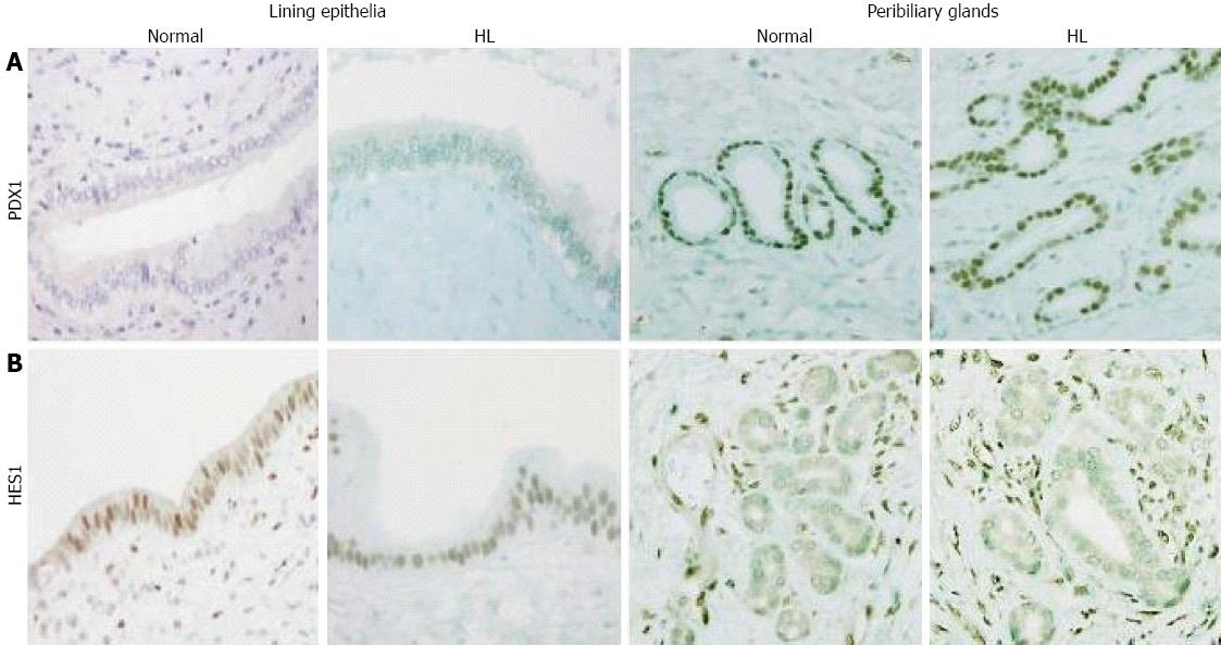
Figure 1 Expression of pancreatic duodenal homeobox factor 1 and hairy and enhancer of split 1.
A: Pancreatic duodenal homeobox factor 1 (PDX1) was not detected in the lining epithelia of hilar bile ducts in normal or hepatolithiatic livers (HL), while it was strongly expressed in peribiliary glands in normal and hepatolithiatic livers. Immunostaining of PDX1 × 200; B: Hairy and enhancer of split 1 (HES1) was frequently expressed in the lining epithelia of hilar bile ducts in normal and hepatolithiatic livers, while its expression in peribiliary glands was infrequent in normal and hepatolithiatic livers. Mesenchymal cells were positive for HES1. Immunostaining of HES1 × 200.
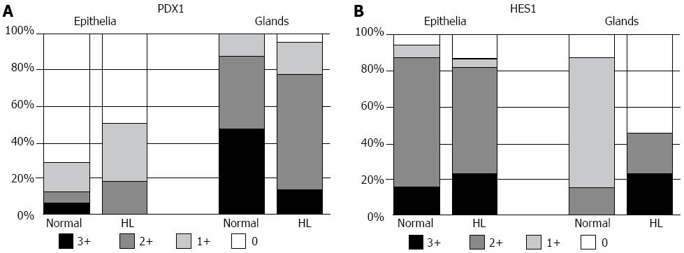
Figure 2 Distribution of pancreatic duodenal homeobox factor 1 and hairy and enhancer of split 1 expression in normal and hepatolithiatic livers.
A: Pancreatic duodenal homeobox factor 1 (PDX1) was more frequently expressed in the peribiliary glands in normal livers (0, 0 case; 1+, 4 cases; 2+, 13 cases; 3+, 15 cases) than in the lining epithelia (0, 23 cases; 1+, 5 cases; 2+, 2 cases; 3+, 2 cases). This distribution pattern was similar in hepatolithiatic livers (HL), too; B: In normal livers, hairy and enhancer of split 1 (HES1) was more frequently expressed in the lining epithelia (0, 2 cases; 1+, 2 cases; 2+, 23 cases; 3+, 5 cases) than in the peribiliary glands (0, 7 cases; 1+, 20 cases; 2+, 5 cases; 3+, 0 case). Although such a distribution pattern was similar in hepatolithiatic livers, HES1 expression in the peribiliary glands was decreased.
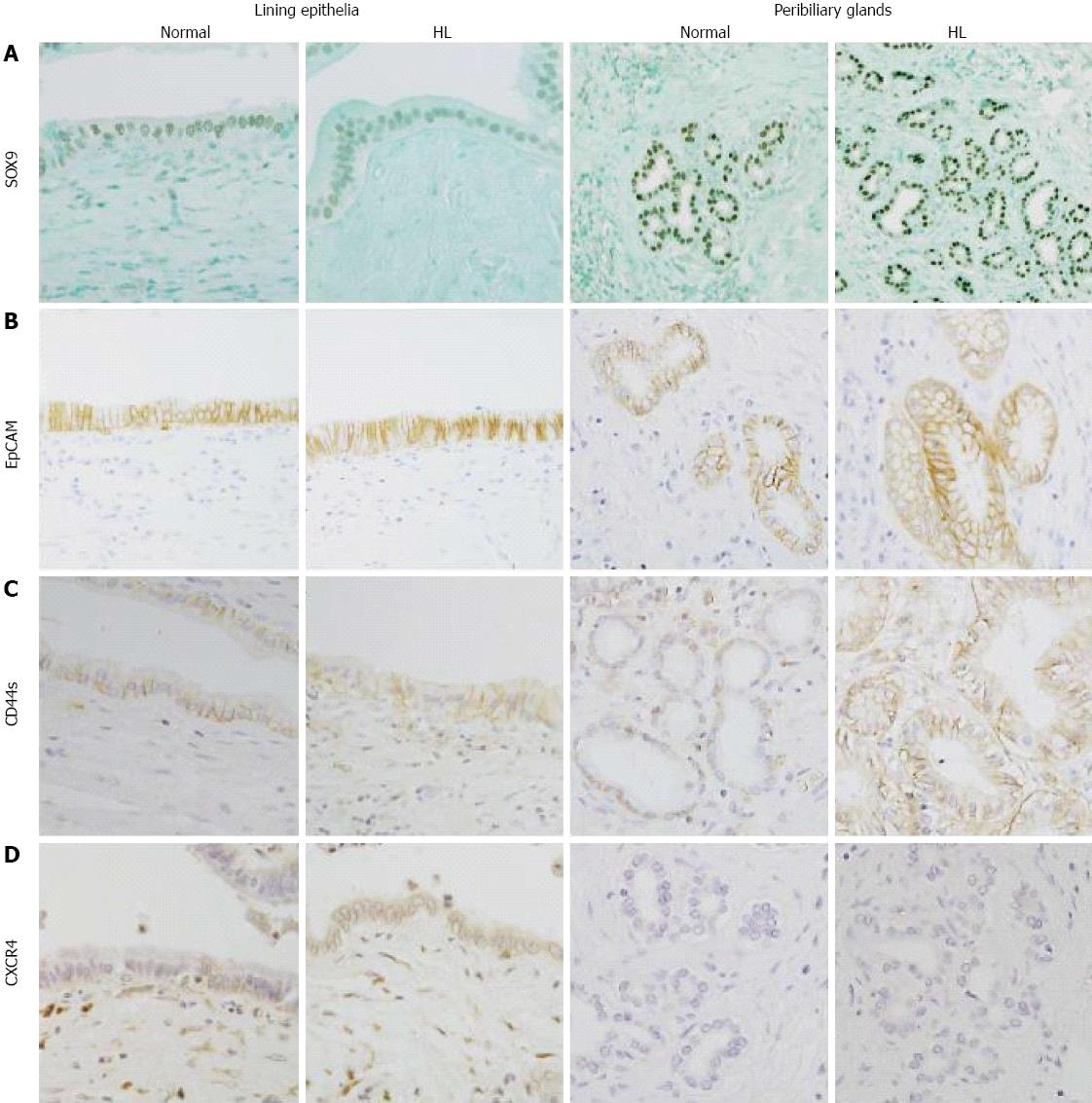
Figure 3 Expression of SOX9, epithelial cell adhesion molecule, CD44s and chemokine receptor type 4.
A: SOX9 was expressed in the nuclei of the lining epithelia of hilar bile ducts in normal livers and of the peribiliary glands in hepatolithiatic livers (HL) (Immunostaining of SOX9, × 200); B: Many of the lining epithelial cells in the hilar bile ducts of normal livers were positive for epithelial cell adhesion molecule (EpCAM) and many of the acini in the peribiliary glands of hepatolithiatic livers were positive for EpCAM (Immunostaining of EpCAM, × 200); C: CD44s was focally expressed in the lining epithelia and peribiliary glands of hepatolithiatic livers (Immunostaining of CD44s, × 200); D: Chemokine receptor type 4 (CXCR4) was focally expressed in the lining epithelia and peribiliary glands of hepatolithiatic livers (Immunostaining of CXCR4, × 200).
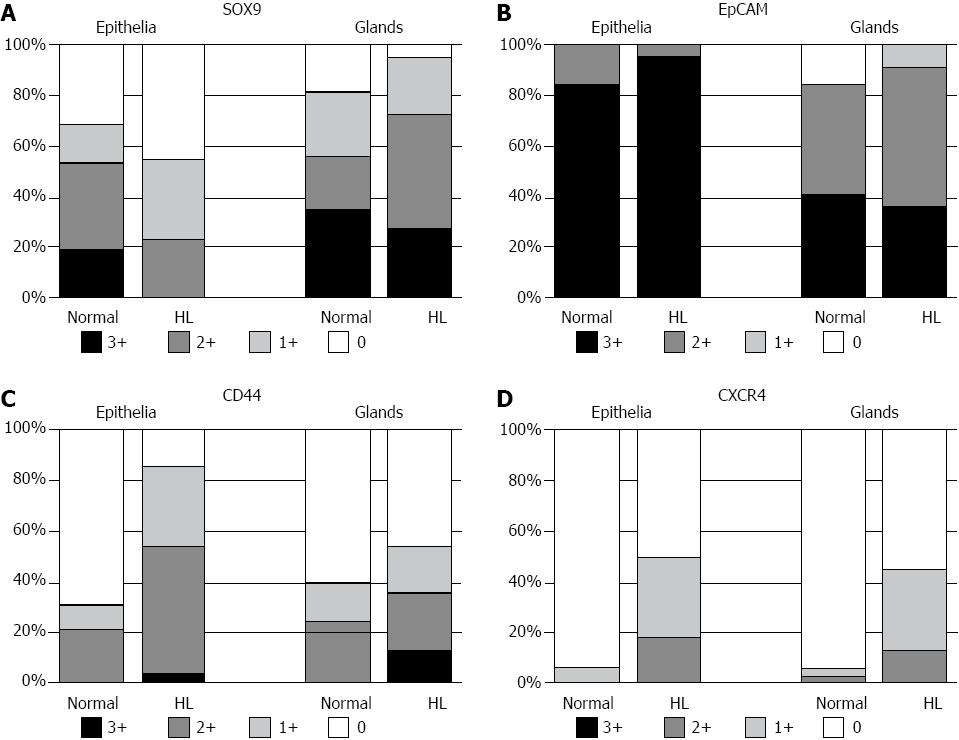
Figure 4 Distribution of SOX9, epithelial cell adhesion molecule, CD44s and chemokine receptor type 4 expression in normal and hepatolithiatic livers.
A: SOX9 was frequently expressed in the lining epithelia and peribiliary glands of normal livers (0, 10 and 6 cases; 1+, 5 and 8 cases; 2+, 11 and 7 cases; 3+, 6 and 11 cases, respectively) and hepatolithiatic livers (0, 10 cases and 1 case; 1+, 7 and 5 cases; 2+, 5 and 10 cases; 3+, 0 and 6 cases, respectively). Its expression was rather higher in the peribiliary glands than in the lining epithelia of hepatolithiatic livers; B: Epithelial cell adhesion molecule (EpCAM) was frequently expressed in the lining epithelia and peribiliary glands of normal livers (0, 0 and 0 case; 1+, 0 case and 5 cases; 2+, 5 and 14 cases; 3+, 27 and 13 cases, respectively) and hepatolithiatic livers (0, 0 and 0 case; 1+, 0 case and 2 cases; 2+, 1 case and 12 cases; 3+, 21 and 8 cases, respectively). Its expression was slightly higher in the lining epithelia of hepatolithiatic livers; C: CD44s expression varied in the lining epithelia and peribiliary glands of normal livers (0, 22 and 19 cases; 1+, 3 and 5 cases; 2+, 6 and 8 cases; 3+, 1 and 0 case, respectively) and hepatolithiatic livers (0, 3 and 10 case; 1+, 7 and 4 cases; 2+, 11 and 5 cases; 3+, 1 case and 3 cases, respectively). The expression of CD44s in the lining epithelia and peribiliary glands was higher in hepatolithiatic livers than in normal livers and a significant difference was observed in the lining epithelia (P < 0.01); D: The expression of chemokine receptor type 4 (CXCR4) was rare in the lining epithelia and peribiliary glands of normal livers (0, 30 and 30 cases; 1+, 0 and 1 case; 2+, 2 cases and 1 case; 3+, 0 and 0 cases, respectively), but was not infrequent in hepatolithiatic livers (0, 11 and 12 cases; 1+, 7 and 7 cases; 2+, 4 and 3 cases; 3+, 0 and 0 case, respectively). A significant difference in its expression in the lining epithelia was observed between normal and hepatolithiatic livers (P < 0.01).
- Citation: Igarashi S, Sato Y, Ren XS, Harada K, Sasaki M, Nakanuma Y. Participation of peribiliary glands in biliary tract pathophysiologies. World J Hepatol 2013; 5(8): 425-432
- URL: https://www.wjgnet.com/1948-5182/full/v5/i8/425.htm
- DOI: https://dx.doi.org/10.4254/wjh.v5.i8.425


