Copyright
©2013 Baishideng Publishing Group Co.
World J Hepatol. Apr 27, 2013; 5(4): 220-225
Published online Apr 27, 2013. doi: 10.4254/wjh.v5.i4.220
Published online Apr 27, 2013. doi: 10.4254/wjh.v5.i4.220
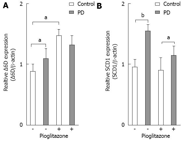
Figure 1 Effect of pioglitazone and MEK inhibition on mRNA expression of Δ6 desaturase and stearoyl-CoA desaturase.
HepG2 cells were incubated for 48 h ± 20 μmol/L pioglitazone with or without 20 μmol/L PD98059 (PD) as indicated. Cell lysates were prepared and analyzed by reverse transcription-polymerase chain reaction for expression levels of Δ6 desaturase (Δ6D) and stearoyl-CoA desaturase (SCD1). Expression of Δ6D (A) and SCD1 (B) in each lysate were quantified and normalized to the amount of β-actin. The mean ± SD of four independent experiments are given. aP < 0.05 and bP < 0.01, Student’s t test, respectively.
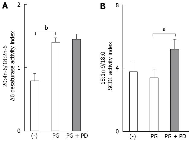
Figure 2 Effect of pioglitazone on derived fatty acid indices of HepG2 human hepatic cells.
A: Δ6 desaturase activity index (20:4n 6/18:2n 6); B: Stearoyl-CoA desaturase 1 (SCD1) activity index (18:1n 9/18:0). Cells were incubated with pioglitazone (PG) (20 μmol/L) and PD98059 (PD; 20 μmol/L) for 48 h. Data are mean ± SD, n = 3. aP < 0.05 and bP < 0.01, Tukey’s test, α = 0.05. 18:0, stearic acid; 18:1n 9, oleic acid; 18:2n 6, linoleic acid; 20:4n 6, arachidonic acid.
- Citation: Saliani N, Darabi M, Yousefi B, Baradaran B, Khaniani MS, Darabi M, Shaaker M, Mehdizadeh A, Naji T, Hashemi M. PPARγ agonist-induced alterations in Δ6-desaturase and stearoyl-CoA desaturase 1: Role of MEK/ERK1/2 pathway. World J Hepatol 2013; 5(4): 220-225
- URL: https://www.wjgnet.com/1948-5182/full/v5/i4/220.htm
- DOI: https://dx.doi.org/10.4254/wjh.v5.i4.220


