Copyright
©2013 Baishideng Publishing Group Co.
World J Hepatol. Mar 27, 2013; 5(3): 156-159
Published online Mar 27, 2013. doi: 10.4254/wjh.v5.i3.156
Published online Mar 27, 2013. doi: 10.4254/wjh.v5.i3.156
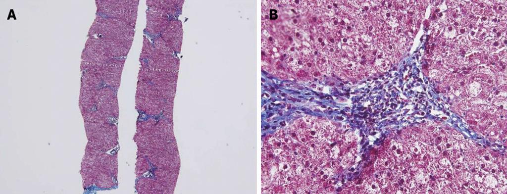
Figure 1 Histological findings.
Biopsy specimen showing mild lobular activity, mild portoperiportal activity and periportal fibrosis with frequent portal to portal bridging fibrosis. A: Masson’s Trichrome stain, 40×; B: Masson’s Trichrome stain, 400×.
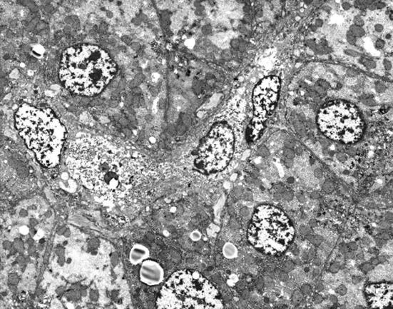
Figure 2 Electron microscopic examination.
There was no evidence of abnormal mitochondria. Some fat vacuoles are found. In the portal area, a few inflammatory cells and collagen depositions are observed (original magnification 2000×).
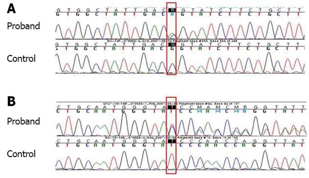
Figure 3 Adenosine triphosphatase 7B sequencing.
A: c.3556G>A, p.G1186S, heterozygote; B: c.4006delA, p.Ile1336TyrfsX57 (reverse complementary sequence, STOP 1392), heterozygote.
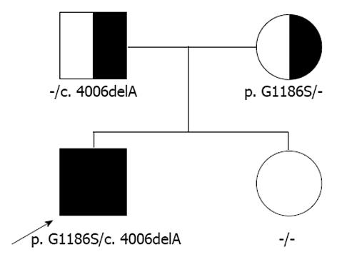
Figure 4 Pedigree.
The patient has a compound heterozygote of p.G1186S and c.4006delA.
- Citation: Kim JW, Kim JH, Seo JK, Ko JS, Chang JY, Yang HR, Kang KH. Genetically confirmed Wilson disease in a 9-month old boy with elevations of aminotransferases. World J Hepatol 2013; 5(3): 156-159
- URL: https://www.wjgnet.com/1948-5182/full/v5/i3/156.htm
- DOI: https://dx.doi.org/10.4254/wjh.v5.i3.156


