Copyright
©2012 Baishideng Publishing Group Co.
World J Hepatol. Dec 27, 2012; 4(12): 412-414
Published online Dec 27, 2012. doi: 10.4254/wjh.v4.i12.412
Published online Dec 27, 2012. doi: 10.4254/wjh.v4.i12.412
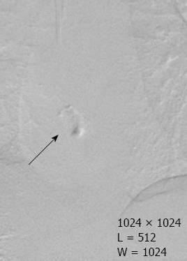
Figure 1 Portal vein embolization material embolism in the right atrial cavity (arrow).
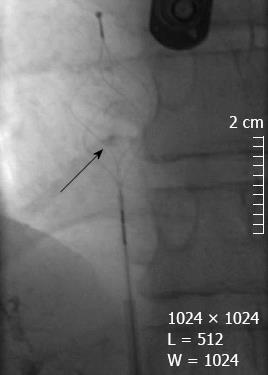
Figure 2 Material withdrawal through the vena cava with a basket catheter (arrow).
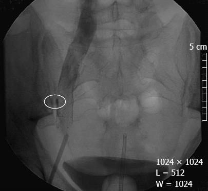
Figure 3 Femoral stent placement.
The stent pressed the portal vein embolization material against the wall of the femoral vein (circle). Note the good venous flow at the end of the procedure.
- Citation: Bouras AF, Truant S, Beregi JP, Sergent G, Delemazure O, Liddo G, Lebuffe G, Zerbib P, Pruvot FR, Boleslawski E. Atrial embolism caused by portal vein embolization: Treatment by percutaneous withdrawal and stenting. World J Hepatol 2012; 4(12): 412-414
- URL: https://www.wjgnet.com/1948-5182/full/v4/i12/412.htm
- DOI: https://dx.doi.org/10.4254/wjh.v4.i12.412


