Copyright
©2011 Baishideng Publishing Group Co.
World J Hepatol. Sep 27, 2011; 3(9): 256-261
Published online Sep 27, 2011. doi: 10.4254/wjh.v3.i9.256
Published online Sep 27, 2011. doi: 10.4254/wjh.v3.i9.256
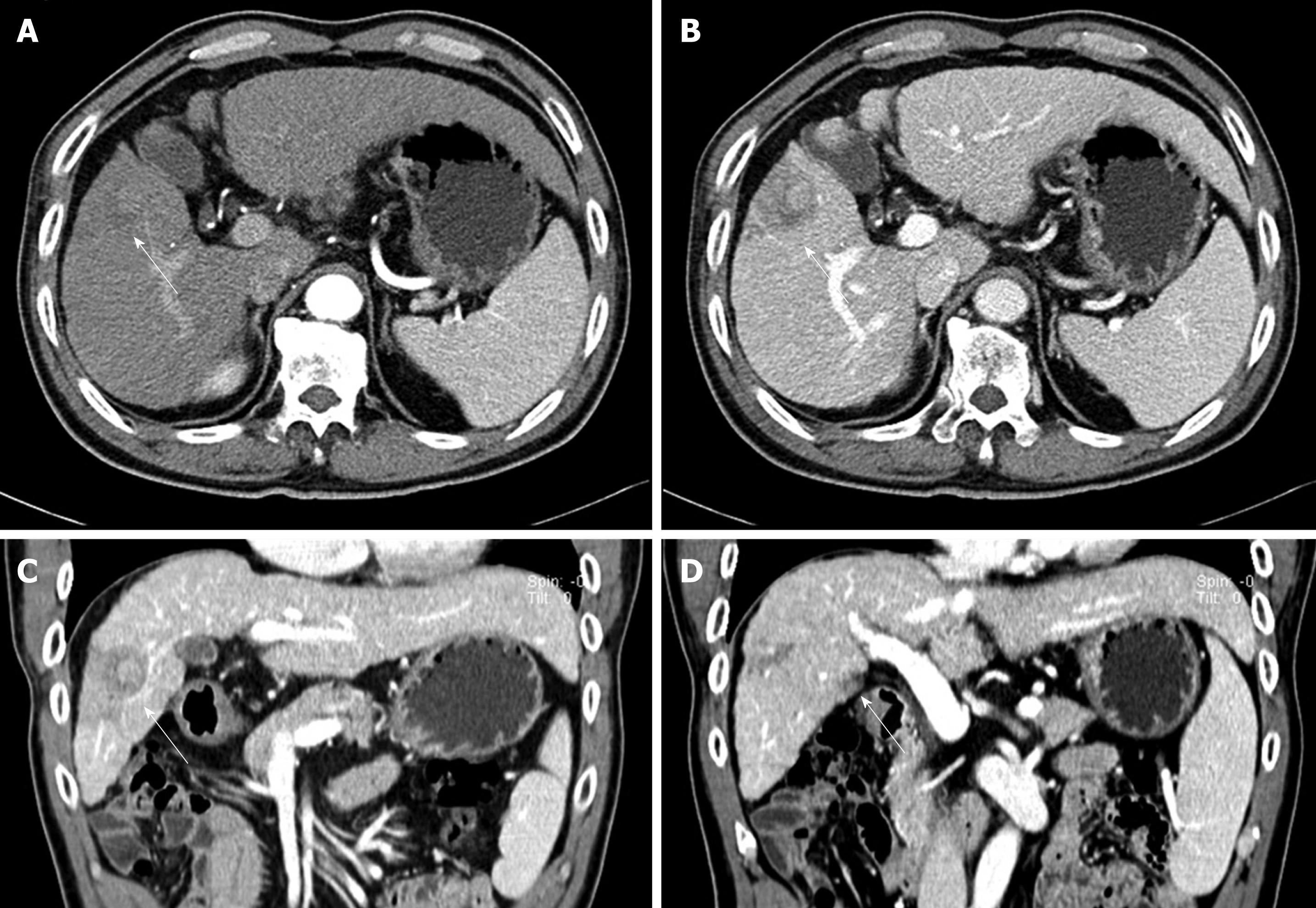
Figure 1 Computed tomography scan results.
A: An ill-defined, low-attenuated lesion was observed in the right anterior segment on late arterial phase image (arrow); B: The lesion was more clearly seen as a low-attenuated, poorly enhanced lesion in the portal phase. Central high-attenuated portion was observed; C: Coronal reformat image in the portal phase, showing the low-attenuated lesion pushing against the segmental portal vein, resulting in its narrowing (arrow); D: Another coronal reformat image in the portal phase, showing a low-attenuated, poorly enhanced lesion in segment V (arrowhead).
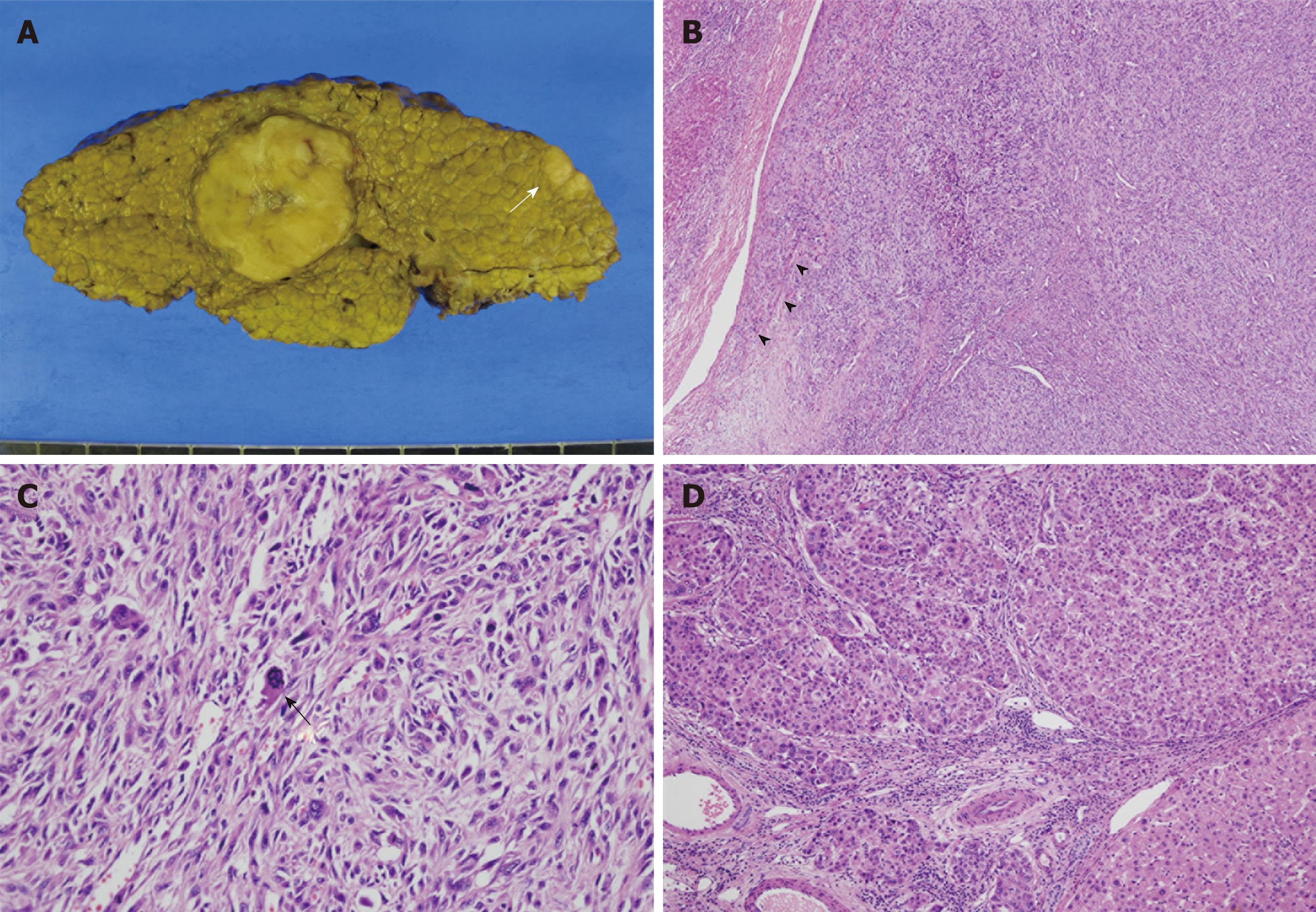
Figure 2 Histopathological findings of the tumors.
A: Gross photography of the resected specimen, showing a large round lobulated mass and a smaller oval-shaped mass (white arrow). Typical multinodular cirrhotic change in background liver was evident; B: Photomicrography of the large round mass, consisting of closely packed spindle cells forming a storiform pattern. Infiltration into the segmental portal vein was also observed (arrowheads) (HE, × 40); C: High power examination, showing pleomorphic spindle cells with bizarre-shaped giant cells, along with numerous mitoses with atypical figures (black arrow) (HE, × 200); D: Photomicrography of the small oval-shaped mass showing polygonal epithelial cells forming trabeculae and round cell nests, consistent with a conventional HCC (HE, × 100).
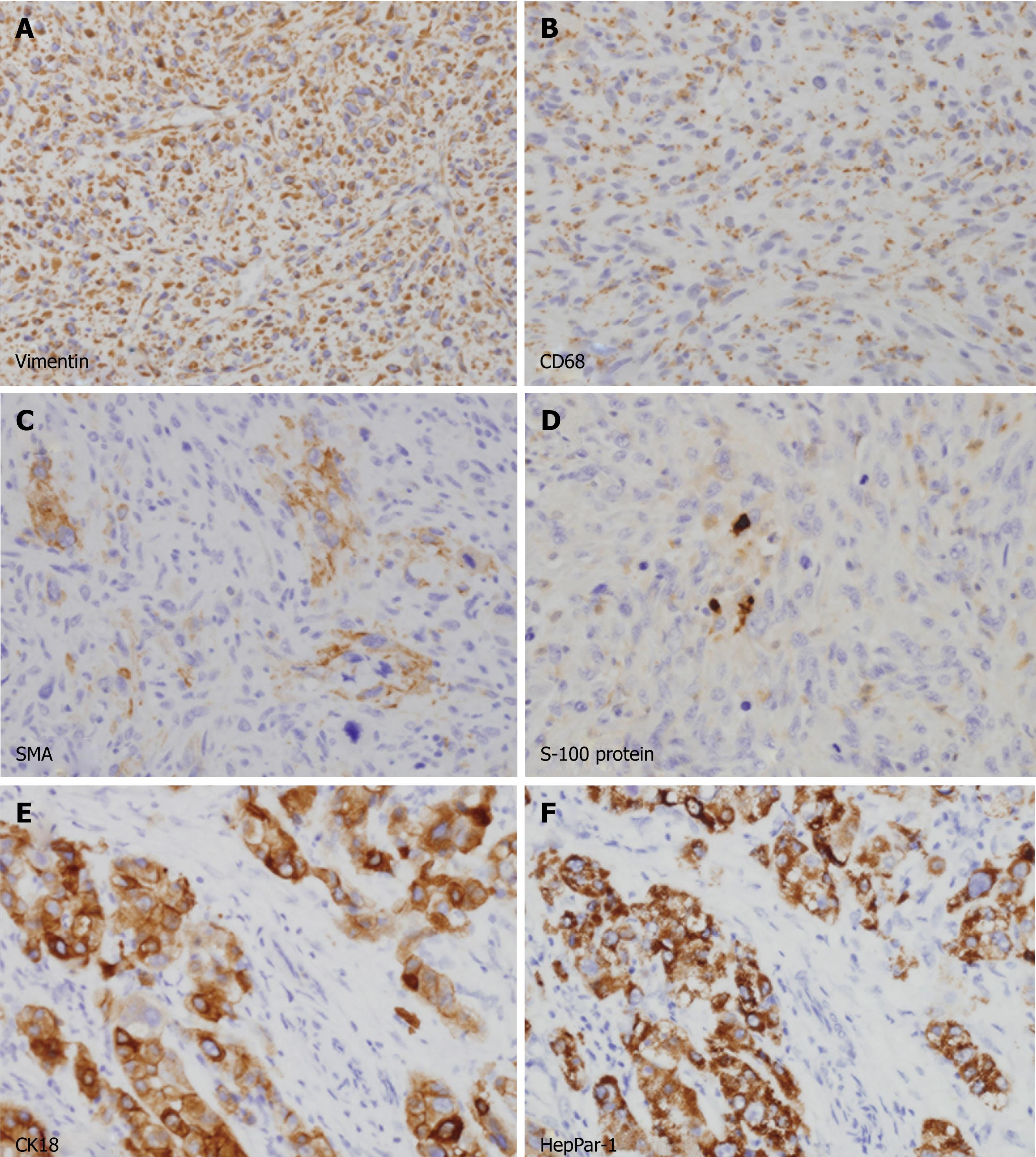
Figure 3 Immunohistochemical analyses of the tumors.
A-D: Spindle cells in the large mass were diffusely immunopositive for vimentin (A) and CD68 (B) in their cytoplasm. A few cells (less than 5%) were also positive for α-smooth muscle actin (C) and S-100 protein (D). All cells were negative for cytokeratins, hepatocyte paraffin-1 antigen (HepPar-1), desmin and myogenic differentiation-1, excluding cells of epithelial and myogenic differentiation (not shown); E, F: Polygonal cells in small mass were strongly immunopositive for cytokeratin 18 (E) and HepPar-1 (F), consistent with a typical hepatocellular carcinoma.
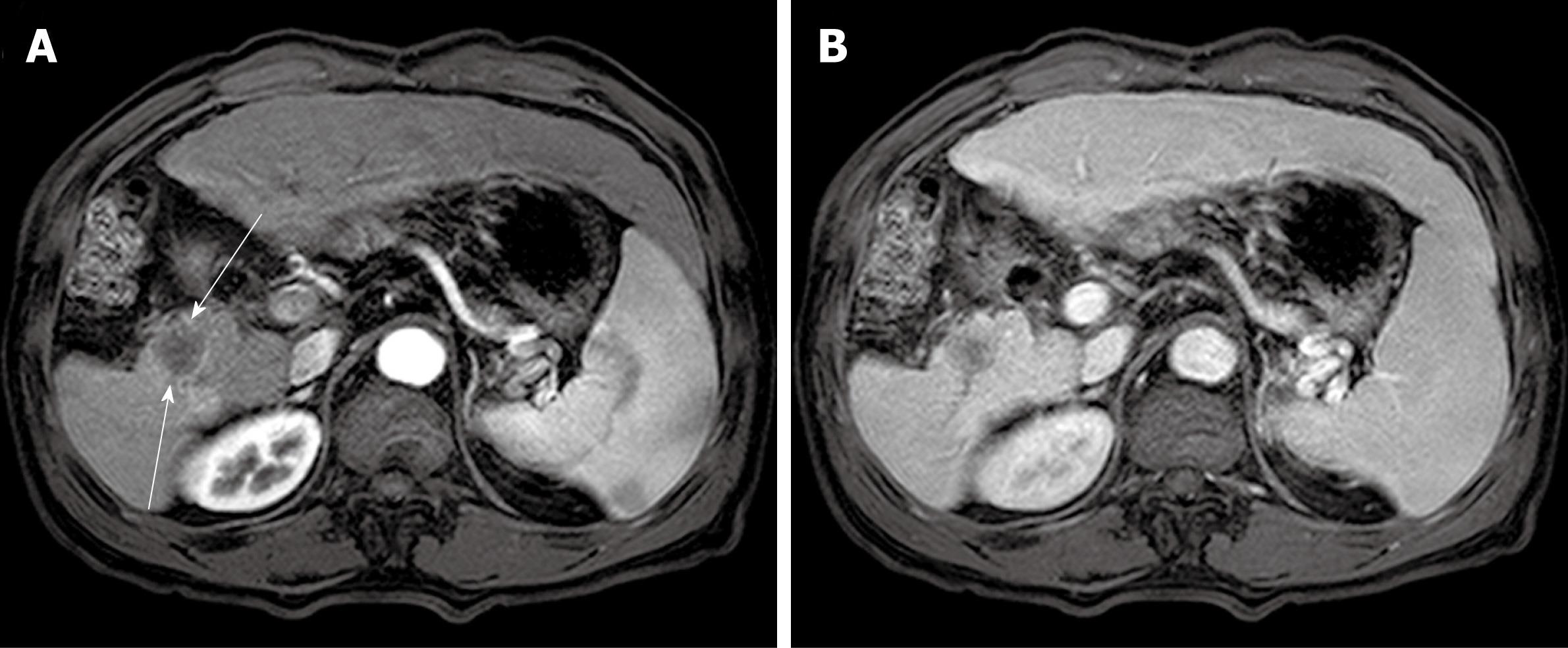
Figure 4 Computed tomography scan at follow-up of twenty-one months.
A: A well demarcated low-attenuated round mass was newly found at the resection margin of right posterior segment (slender arrows). This mass was not significantly enhanced in late arterial phase; B: This mass was also not well enhanced at portal phase, which is similar pattern with the previous malignant fibrous histiocytoma.
- Citation: Hwang HS, Ha ND, Jeong YK, Suh JH, Choi HJ, Kim YM, Cha HJ. Simultaneous occurrence of malignant fibrous histiocytoma and hepatocellular carcinoma in cirrhotic liver: A case report. World J Hepatol 2011; 3(9): 256-261
- URL: https://www.wjgnet.com/1948-5182/full/v3/i9/256.htm
- DOI: https://dx.doi.org/10.4254/wjh.v3.i9.256


