Copyright
©2010 Baishideng Publishing Group Co.
World J Hepatol. Sep 27, 2010; 2(9): 354-357
Published online Sep 27, 2010. doi: 10.4254/wjh.v2.i9.354
Published online Sep 27, 2010. doi: 10.4254/wjh.v2.i9.354
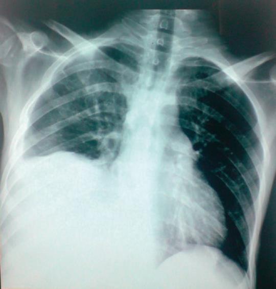
Figure 1 Chest X-ray showing a right-sided sub diaphragmatic pathology as the right hemi-diaphragm is raised and the costophrenic angle is blunted.
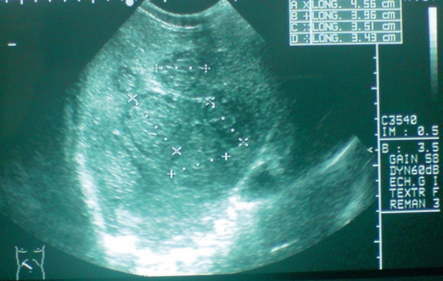
Figure 2 Ultrasonography of the abdomen revealing heterogeneous hypo-echoic lesions in the right lobe of the liver suggestive of a multiseptate abscess.
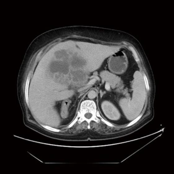
Figure 3 Computerized tomography of abdomen showing the multiseptate abscess in the right lobe of liver.
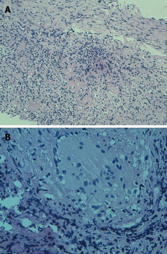
Figure 4 Histological examination of the abscess wall showing a Liver epithelioid granuloma with giant-cell and foci of caseous necrosis.
A: HES × 10; B: HES × 30.
- Citation: Hassani KIM, Ousadden A, Ankouz A, Mazaz K, Taleb KA. Isolated liver tuberculosis abscess in a patient without immunodeficiency: A case report. World J Hepatol 2010; 2(9): 354-357
- URL: https://www.wjgnet.com/1948-5182/full/v2/i9/354.htm
- DOI: https://dx.doi.org/10.4254/wjh.v2.i9.354


