Copyright
©The Author(s) 2025.
World J Hepatol. Aug 27, 2025; 17(8): 109093
Published online Aug 27, 2025. doi: 10.4254/wjh.v17.i8.109093
Published online Aug 27, 2025. doi: 10.4254/wjh.v17.i8.109093
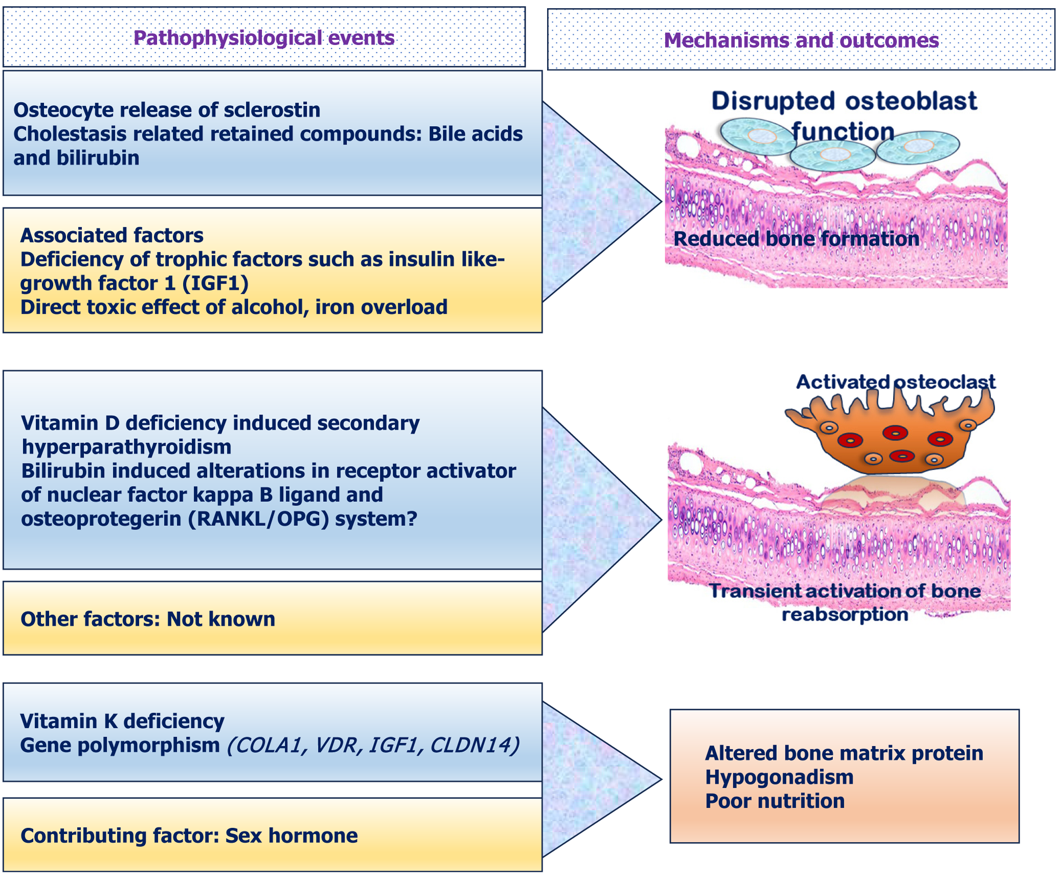
Figure 1 Outline of pathogenesis of hepatic osteodystrophy: Reduced osteoblast function, activated functions of osteoclasts and other factors.
IGF-1: Insulin-like growth factor 1; OPG: Osteoprotegerin; RANKL: Receptor activator of nuclear factor kappa-B ligand.
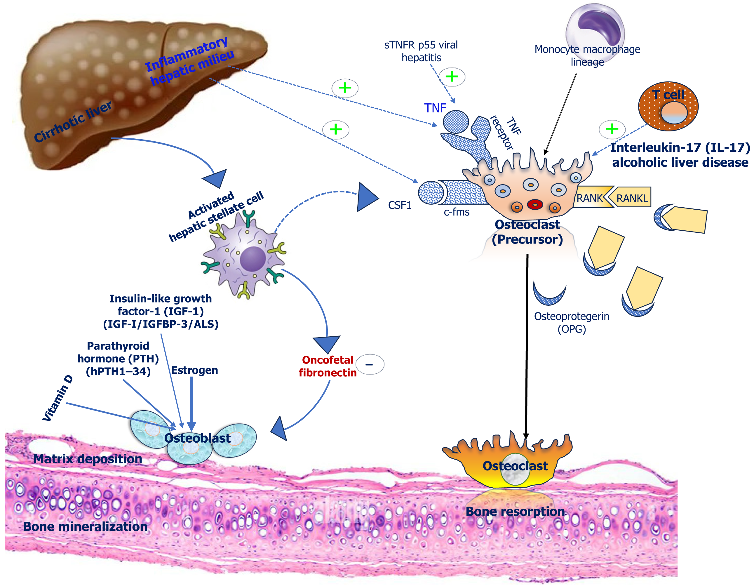
Figure 2 Role of inflammatory cytokines and hormonal factors in the pathogenesis of hepatic osteodystrophy (Solid arrows display known pathways, while interrupted arrows suggest pathways with little evidence).
IGF-1: Insulin-like growth factor 1; OPG: Osteoprotegerin; PTH: Parathyroid hormone; TNF: Tumor necrosis factor.
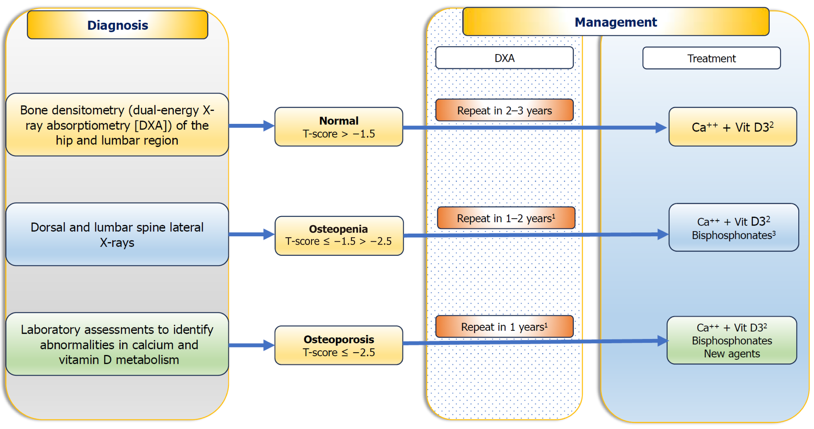
Figure 3 Diagnosis and treatment of bone disorders among patients with chronic liver disease[80].
1According to the severity of liver disease and cholestasis, and in patients taking steroids; 2Calcium (1000–1500 mg/day) and vitamin D (400–800 IU/day) are necessary to maintain normal levels; 3Depending on additional risk factors. DXA: Dual energy X-ray absorptiometry.
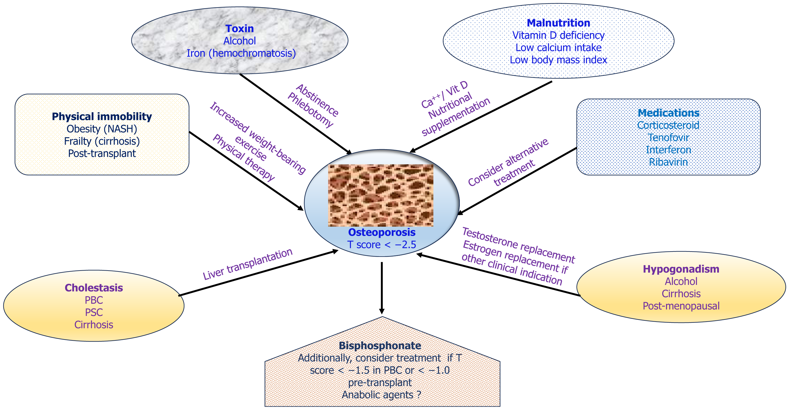
Figure 4 Schematic diagram of contributing factors to the development of osteoporosis in chronic liver disease, preventive interventions, and treatment of osteoporosis[1].
PBC: Primary biliary cholangitis; PSC: Primary sclerosing cholangitis; NASH: Non-alcoholic steatohepatitis.
- Citation: Pramanik S, Palui R, Ray S. Hepatic osteodystrophy: An underrecognized metabolic bone disease. World J Hepatol 2025; 17(8): 109093
- URL: https://www.wjgnet.com/1948-5182/full/v17/i8/109093.htm
- DOI: https://dx.doi.org/10.4254/wjh.v17.i8.109093


