Copyright
©The Author(s) 2025.
World J Hepatol. Jul 27, 2025; 17(7): 106253
Published online Jul 27, 2025. doi: 10.4254/wjh.v17.i7.106253
Published online Jul 27, 2025. doi: 10.4254/wjh.v17.i7.106253
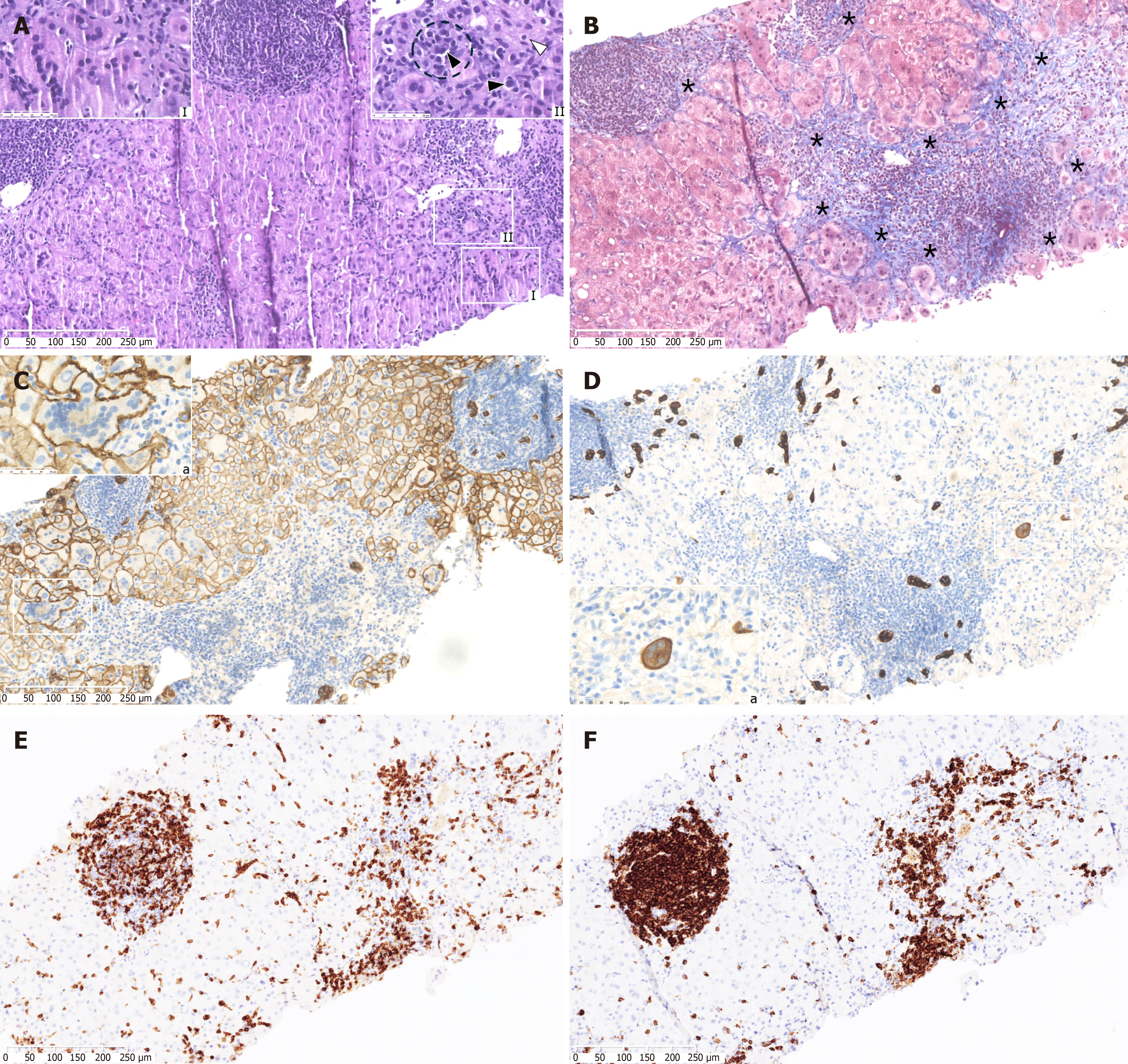
Figure 1 Histopathological features.
A: Many giant multinucleated hepatocytes were frequently observed in both periportal and pericentral lobule areas (I). Mononuclear cells together with typical clustered plasma cells (black arrowhead and dashed circle) infiltrate enlarged and edematous portal tract and spill over in the adjacent lobule surrounding hepatocytes (II). Emperipolesis was also detected (white arrowhead) (II); B: Groups of hepatocytes were surrounded by periportal fibrosis in the Trichrome staining (*); C: Immunohistochemistry for E-cadherin better shows the boundaries of giant hepatocytes (I); D: Ductular reaction was detected together with scattered hepatobiliary cells cytokeratin 7-positive, some of which were multinucleated featured (I); E and F: Mononuclear cell infiltrates mainly located in portal tract are mixed, showing both T-cell and B-cell marker expression and excluding infiltration of the liver by chronic lymphocytic leukemia cells (cluster of differentiation 3 [CD3] and CD20, respectively in E and F panels). Optical microscope 100 ×; High-power fields 600 ×.
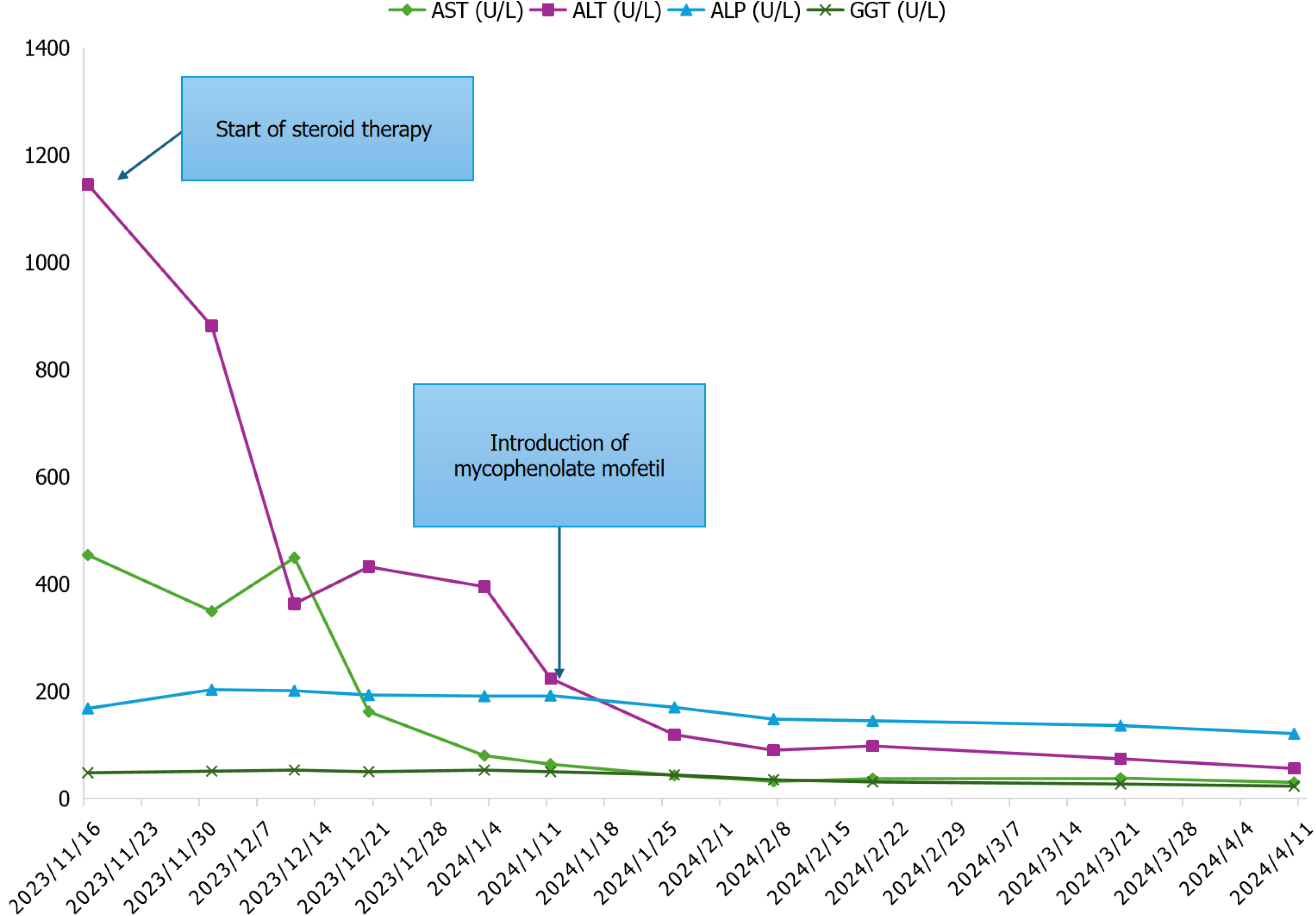
Figure 2 Liver tests during therapy.
The graph shows the trend of liver enzymes over time and demonstrates how the addition of mycophenolate mofetil to the therapy led to a complete biochemical response. ALP: Alkaline phosphatase; ALT: Alanine aminotransferase; AST: Aspartate aminotransferase; GGT: Gamma-glutamyltransferase.
- Citation: Giacomelli M, Carotti S, Vozella F, Pagliei F, Taffon C, Baiocchini A, Gambaro FL, Picardi A, Vespasiani-Gentilucci U, Galati G. Autoimmune hepatitis with syncytial giant cells in chronic lymphocytic leukemia: A case report and literature review. World J Hepatol 2025; 17(7): 106253
- URL: https://www.wjgnet.com/1948-5182/full/v17/i7/106253.htm
- DOI: https://dx.doi.org/10.4254/wjh.v17.i7.106253


