Copyright
©The Author(s) 2025.
World J Hepatol. Mar 27, 2025; 17(3): 103854
Published online Mar 27, 2025. doi: 10.4254/wjh.v17.i3.103854
Published online Mar 27, 2025. doi: 10.4254/wjh.v17.i3.103854
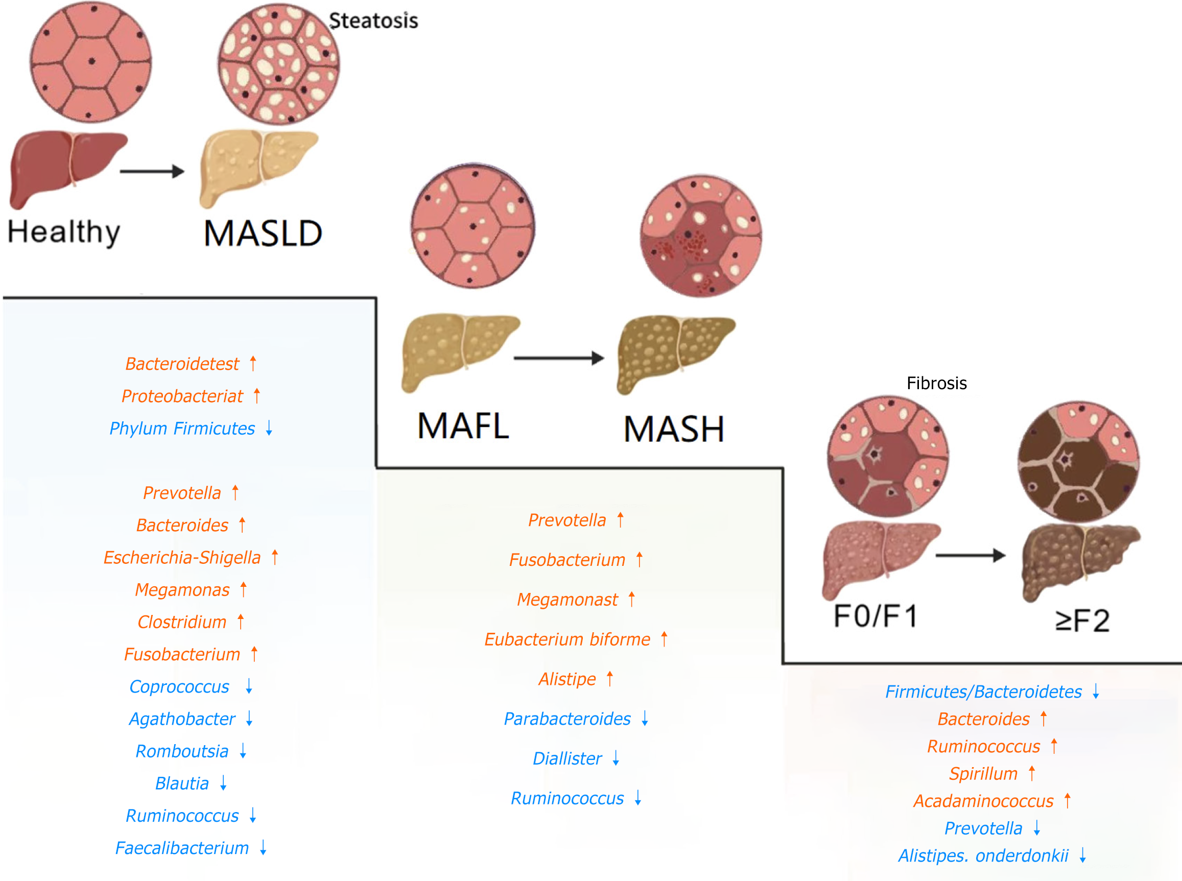
Figure 1 The changes in healthy livers, livers at different stages of diseases, and local liver cells, as well as the changes in some gut microbiota in the pathological states of the liver at various stages.
MASLD: Metabolic dysfunction-associated steatotic liver disease; MAFL: Metabolic dysfunction-associated fatty liver; MASH: Metabolic dysfunction-associated steatohepatitis. Created from BIOGDP.com[93].
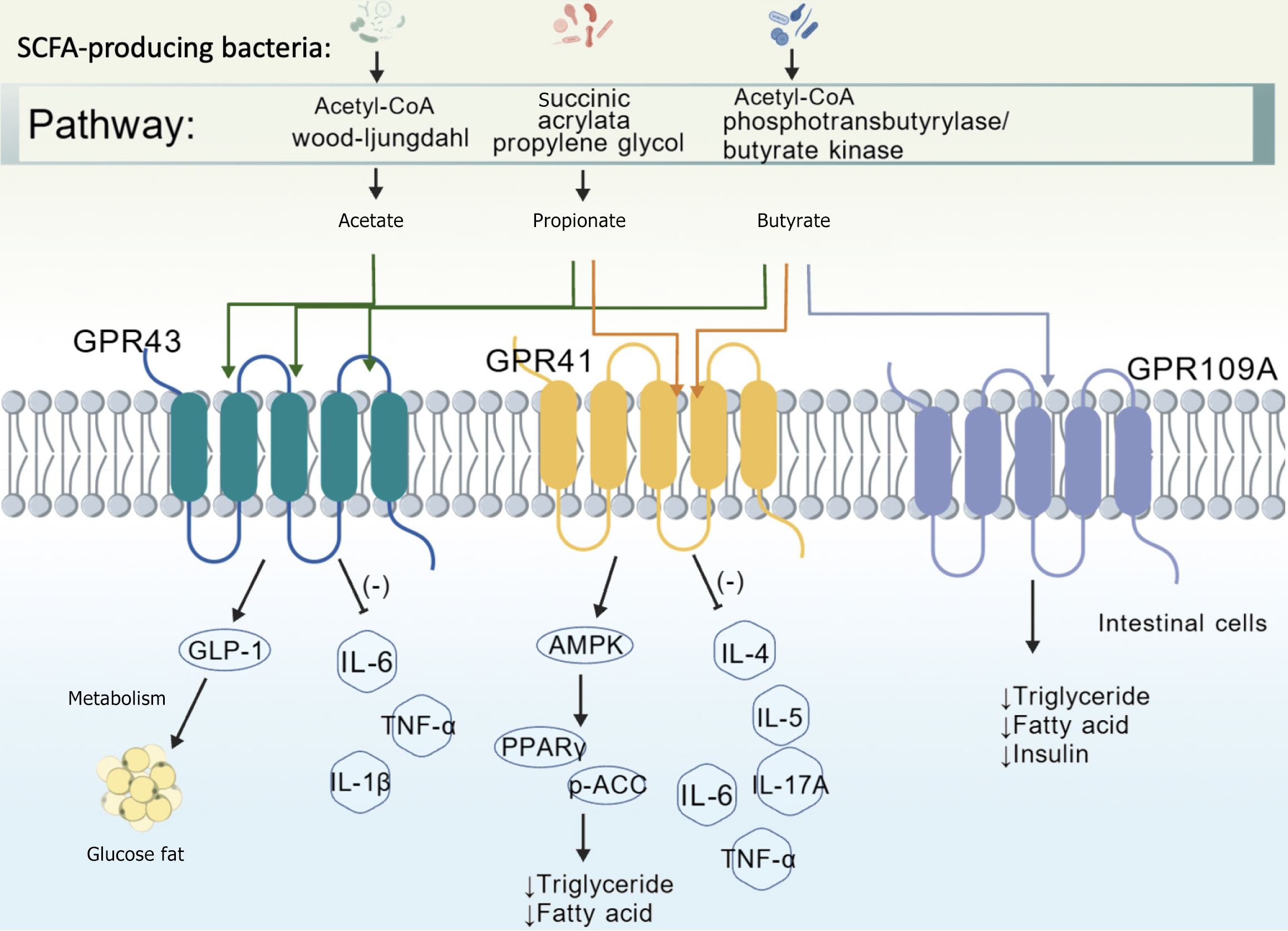
Figure 2 The therapeutic effects of different gut microbiota on diseases through the production of short-chain fatty acids via different pathways and their binding to GPR43, GPR41 and GPR109A receptors.
SCFA: Short-chain fatty acid. Created from BIOGDP.com[93].
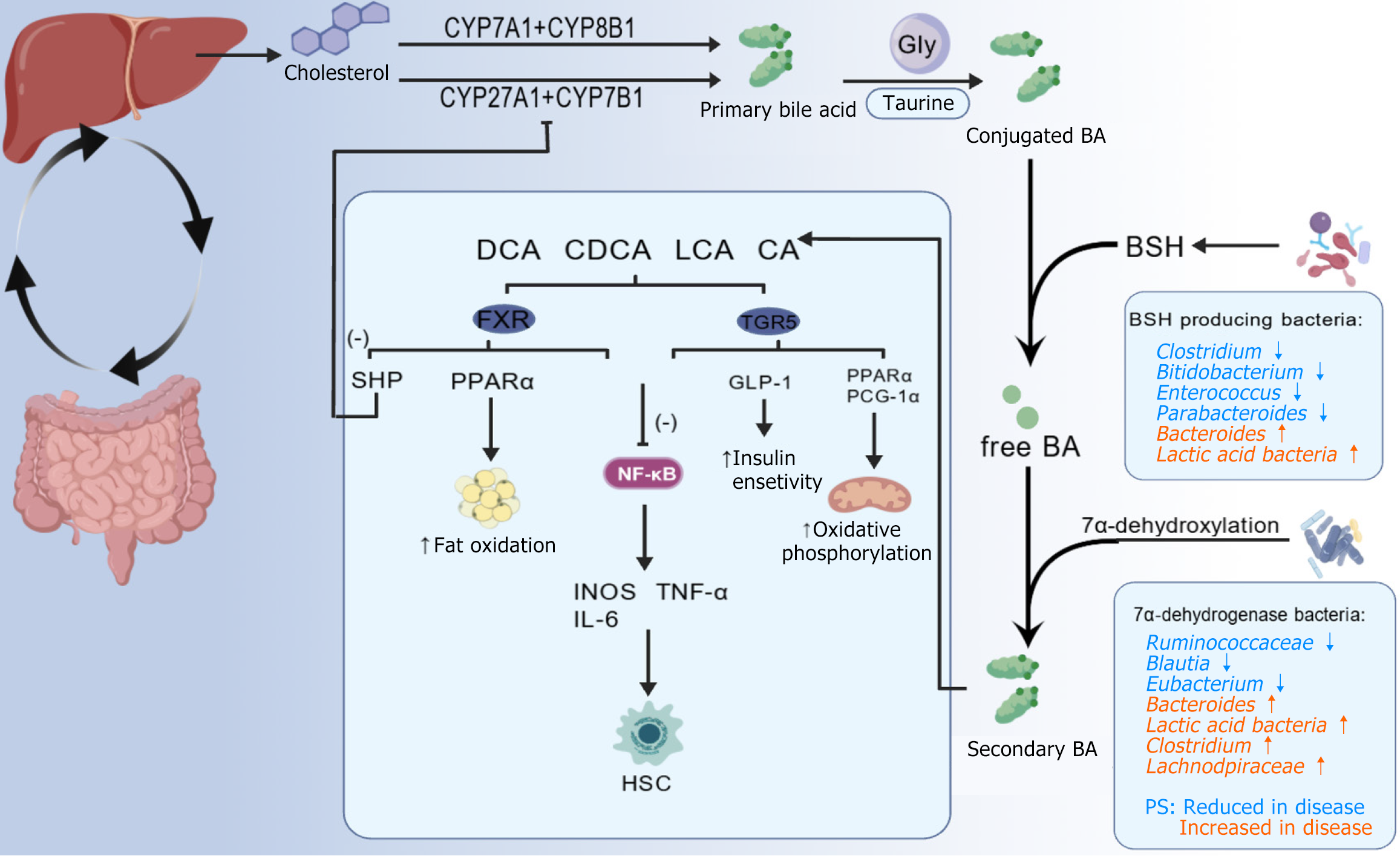
Figure 3 In the enterohepatic circulation, cholesterol within the liver is metabolized into bile acids via two distinct pathways.
Influenced by the gut microbiota, primary bile acids are further transformed into secondary bile acids, which include cholic acid (CA), chenodeoxycholic acid, lithocholic acid, and cholic acid. These secondary bile acids can activate the farnesoid X receptor and TGR5 receptor, thereby exerting a disease - alleviating effect. DCA: Deoxycholic acid; CDCA: Chenodeoxycholic acid; LCA: Lithocholic acid; CA: Cholic acid; BA: Bile acid; BSH: Bile salt hydrolase; HSC: Hepatic stellate cell. Created from BIOGDP.com[93].
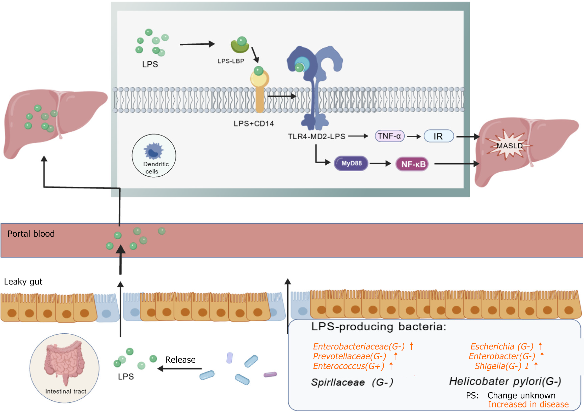
Figure 4 In the intestine, lipopolysaccharide-producing microorganisms spread through the intestinal leakage into the portal vein circulation and then reach the liver, where they release lipopolysaccharide.
By binding to the TLR4-MD2 receptor, this process exacerbates the occurrence of metabolic dysfunction-associated steatotic liver disease. LPS: Lipopolysaccharide; LBP: Lipopolysaccharide-binding protein; IR: Insulin resistance; MASLD: Metabolic dysfunction-associated steatotic liver disease. Created from BIOGDP.com[93].
- Citation: Shu JZ, Huang YH, He XH, Liu FY, Liang QQ, Yong XT, Xie YF. Gut microbiota differences, metabolite changes, and disease intervention during metabolic - dysfunction - related fatty liver progression. World J Hepatol 2025; 17(3): 103854
- URL: https://www.wjgnet.com/1948-5182/full/v17/i3/103854.htm
- DOI: https://dx.doi.org/10.4254/wjh.v17.i3.103854


