Copyright
©The Author(s) 2025.
World J Hepatol. Feb 27, 2025; 17(2): 103299
Published online Feb 27, 2025. doi: 10.4254/wjh.v17.i2.103299
Published online Feb 27, 2025. doi: 10.4254/wjh.v17.i2.103299
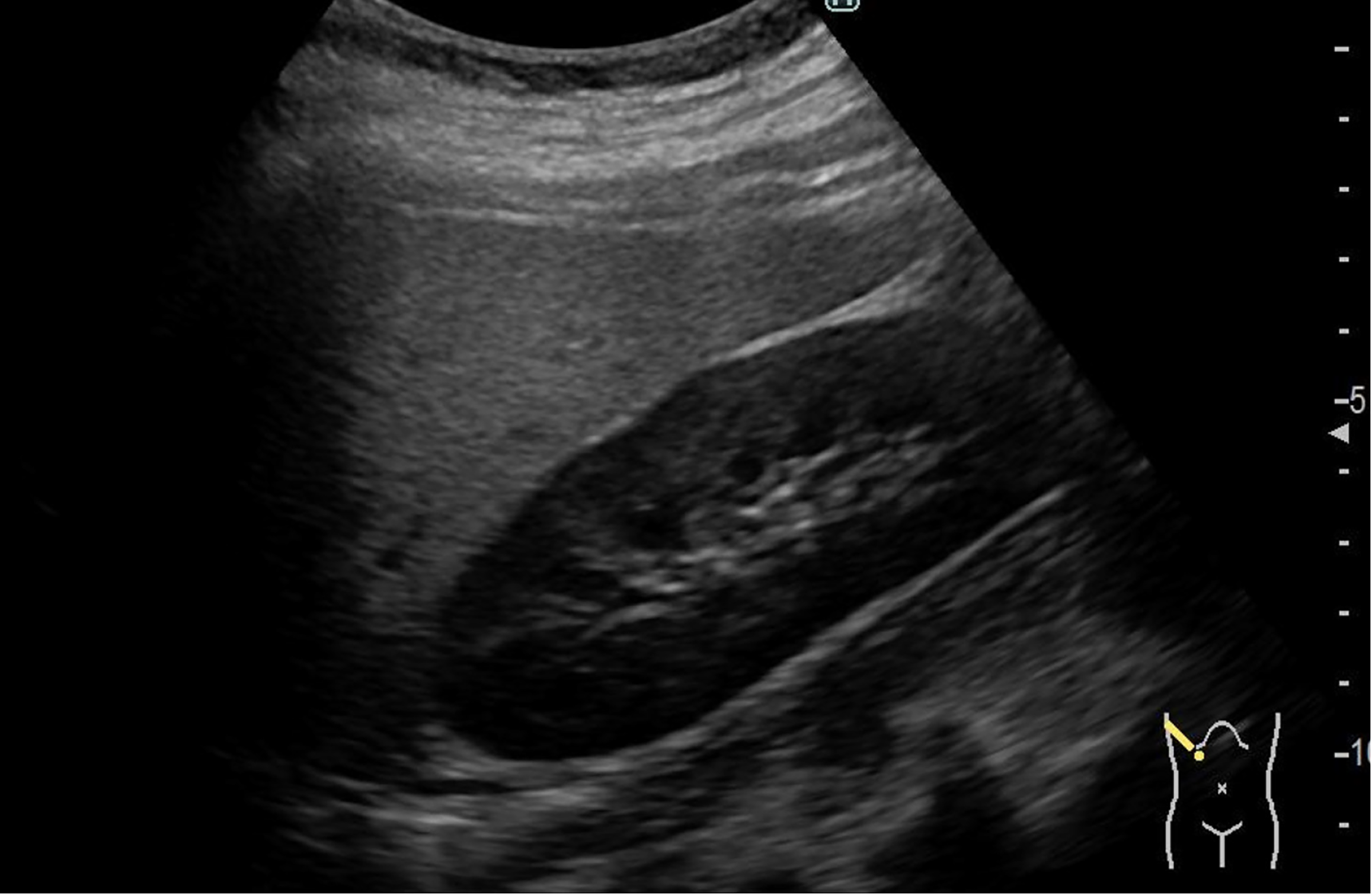
Figure 1 Ultrasound findings.
Ultrasound examination showed no space-occupying lesions in the liver. However, deep attenuation and liver–kidney contrast were observed, suggesting fatty liver.
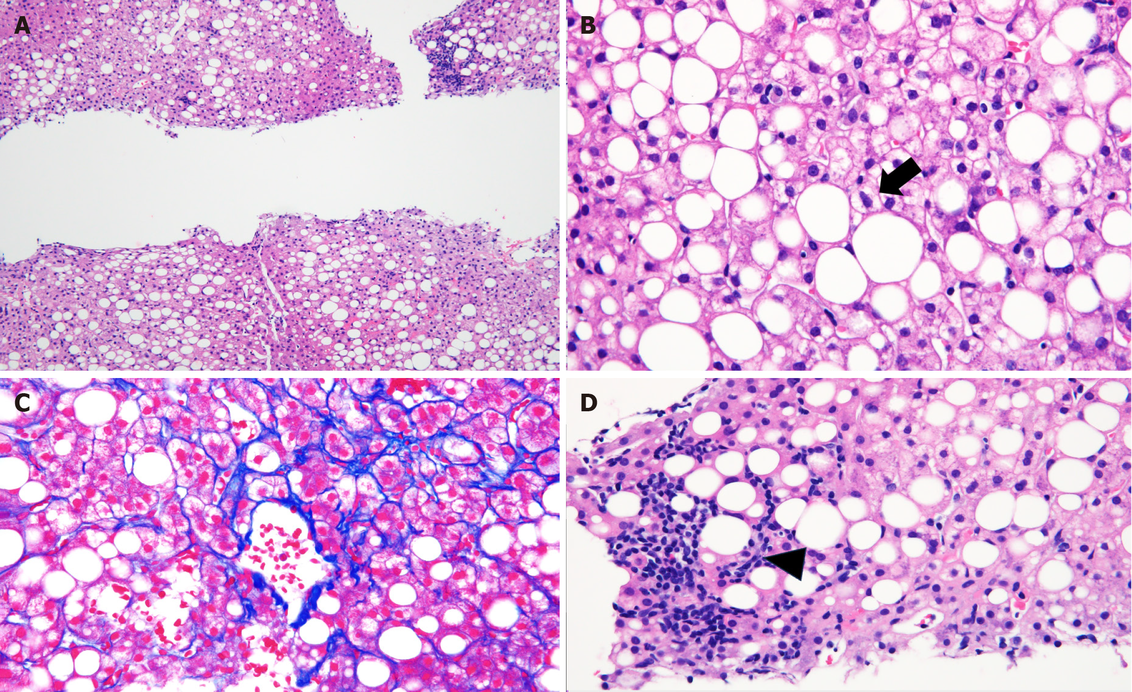
Figure 2 Pathological findings.
A: More than 50% of the hepatocytes showed mixed macro and microvesicular steatosis (hematoxylin-eosin stain, low magnification); B: Some hepatocytes show hepatocellular ballooning (hematoxylin-eosin stain, high magnification); C: Fibrosis around the central veins and hepatocytes (Azan stain, high magnification); D: Lymphocytic infiltration within the hepatic lobules (hematoxylin-eosin stain, high magnification).
- Citation: Miyamoto K, Kondo S, Kondo T, Ishikawa R, Tani R, Inoue T, Matsunaga K, Minamino T, Kusaka T. Pathological features of non-alcoholic steatohepatitis in a pediatric patient with heterozygous familial hypobetalipoproteinemia: A case report. World J Hepatol 2025; 17(2): 103299
- URL: https://www.wjgnet.com/1948-5182/full/v17/i2/103299.htm
- DOI: https://dx.doi.org/10.4254/wjh.v17.i2.103299


