Copyright
©The Author(s) 2024.
World J Hepatol. Feb 27, 2024; 16(2): 279-285
Published online Feb 27, 2024. doi: 10.4254/wjh.v16.i2.279
Published online Feb 27, 2024. doi: 10.4254/wjh.v16.i2.279
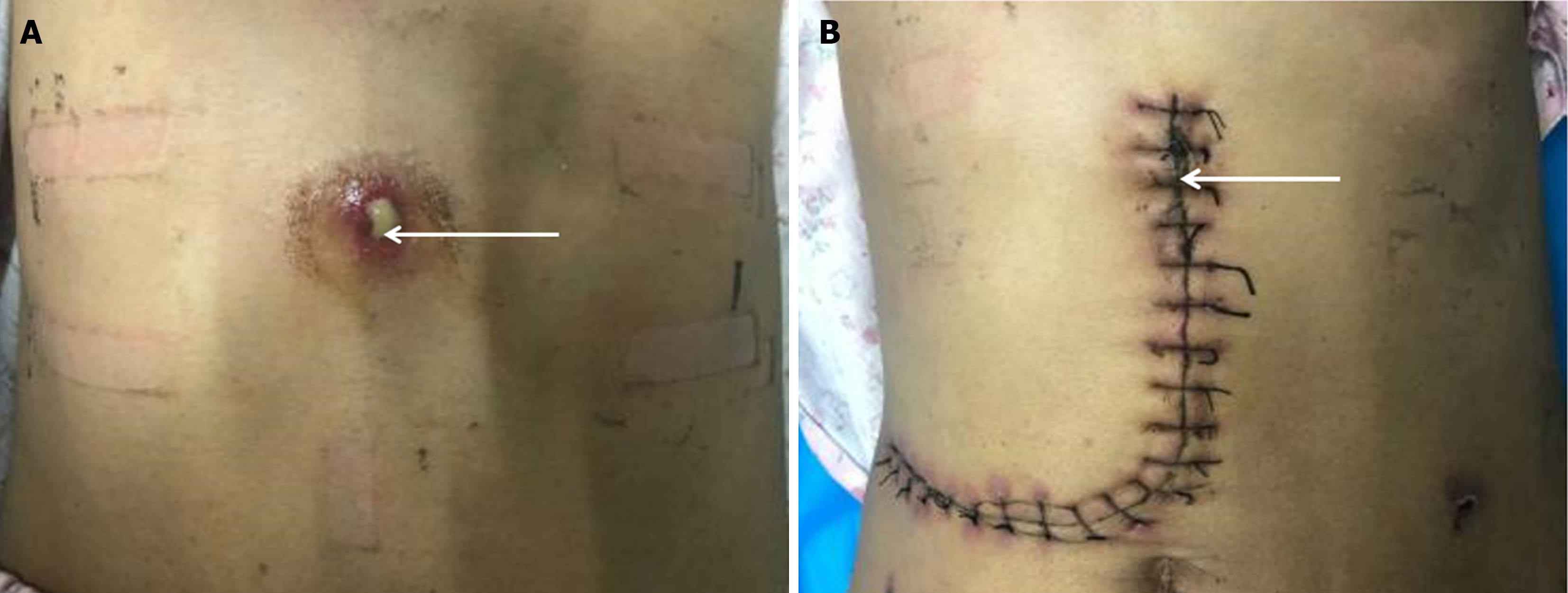
Figure 1 Preoperative lesions and postoperative incisions.
A: Preoperative site of the patient's abdominal wall abscess; B: Postoperative abdominal wall abscess site. Note: The white arrows indicates the site of the abdominal wall abscess sinus tract.
Figure 2 Pre- and postoperative imaging.
A: Preoperative computed tomography (CT) images; B: Preoperative CT images; C: Postoperative CT images. Note: The white arrows indicate the site of the abdominal wall abscess sinus tract, the blue arrows indicate the site of the hepatic alveolar echinococcosis lesion, and the yellow arrows indicates the site of the hepatic cystic echinococcosis lesion.
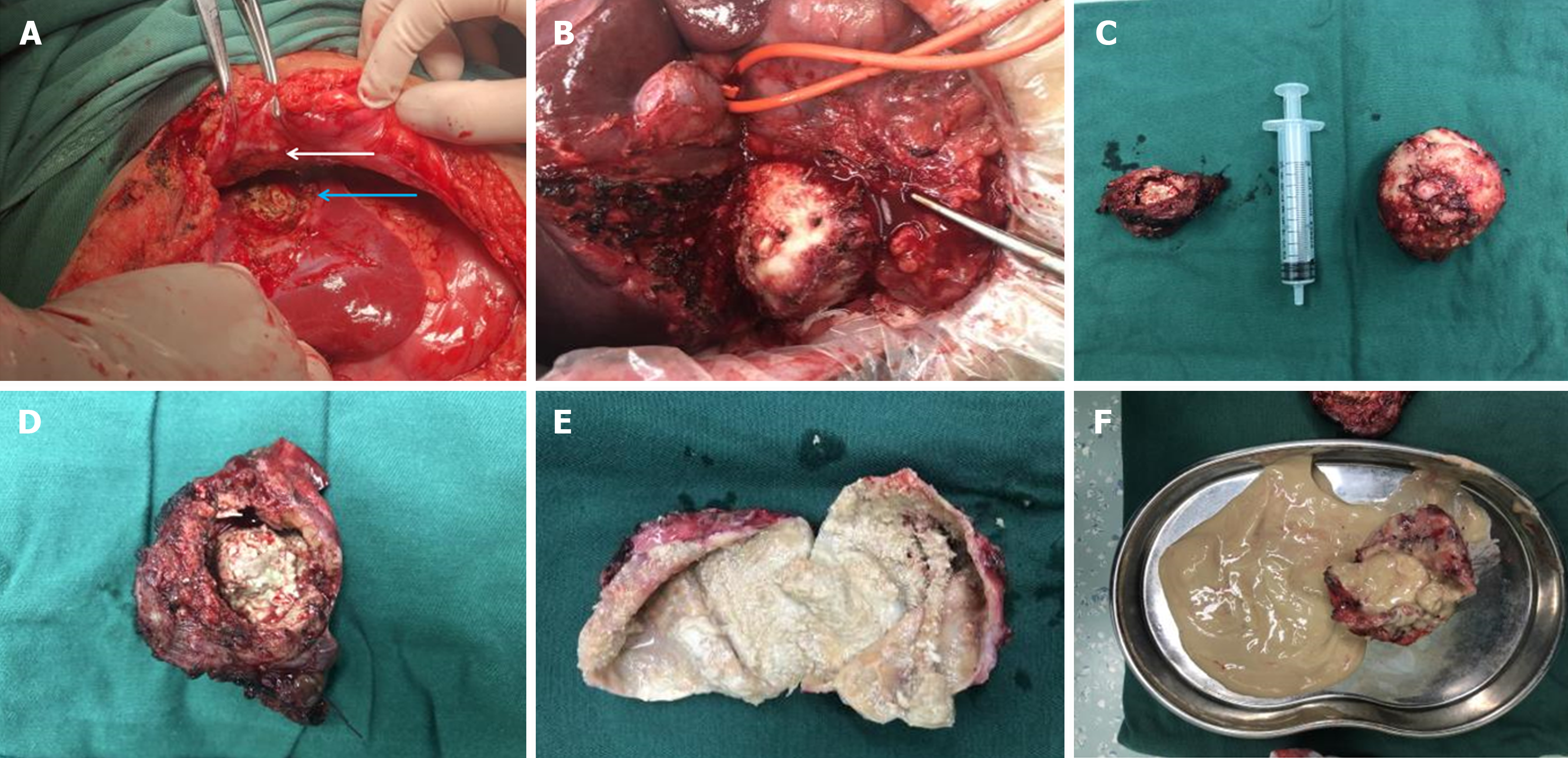
Figure 3 Intraoperative pathology specimens.
A and B: Intraoperative visible lesions; C: Intraoperative excision of pathologic specimens (hepatic cystic echinococcosis and hepatic alveolar echinococcosis); D: Intraoperative excision of pathologic specimens (hepatic alveolar echinococcosis); E: Intraoperative excision of pathologic specimens (hepatic cystic echinococcosis); F: Intraoperative excision of pathologic specimens. Note: The white arrows indicate the site of the abdominal wall abscess sinus tract, and the blue arrows indicate the site of the hepatic alveolar echinococcosis lesion.
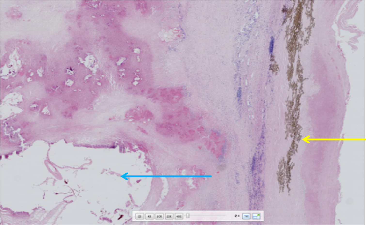
Figure 4
Postoperative pathology slides Histopathological examination by hemotoxylin-eosin staining (200 ×) fibrous connective tissue proliferation and inflammatory cell infiltration are seen around the blue arrow vesicles, forming nodules of varying sizes (alveolar echinococcosis) yellow arrow laminar-like structures are clearly visible (cystic echinococcosis).
- Citation: Wang MM, An XQ, Chai JP, Yang JY, A JD, A XR. Coinfection with hepatic cystic and alveolar echinococcosis with abdominal wall abscess and sinus tract formation: A case report. World J Hepatol 2024; 16(2): 279-285
- URL: https://www.wjgnet.com/1948-5182/full/v16/i2/279.htm
- DOI: https://dx.doi.org/10.4254/wjh.v16.i2.279


